


















Lesson 10 of 52 · Organization of the Body
Tissues, membranes and the skin
Learn the four primary tissues, how epithelium is named, the main connective tissues, tissue repair, body membranes and the layers of the skin.
If cells are the bricks, tissues are the walls: groups of similar cells and the material between them, arranged to do one kind of work. Over a century of microscope study has sorted the human body's many tissues into just four primary types: epithelial, connective, muscle and nervous. Every organ you will meet in the rest of the course is a combination of these four. The study of tissues is called histology, and it is the bridge between cell biology and whole-organ anatomy.
In this lesson you will learn how to recognize each tissue and connect its structure to its job, then see how tissues form the body's membranes and the skin, your largest organ.
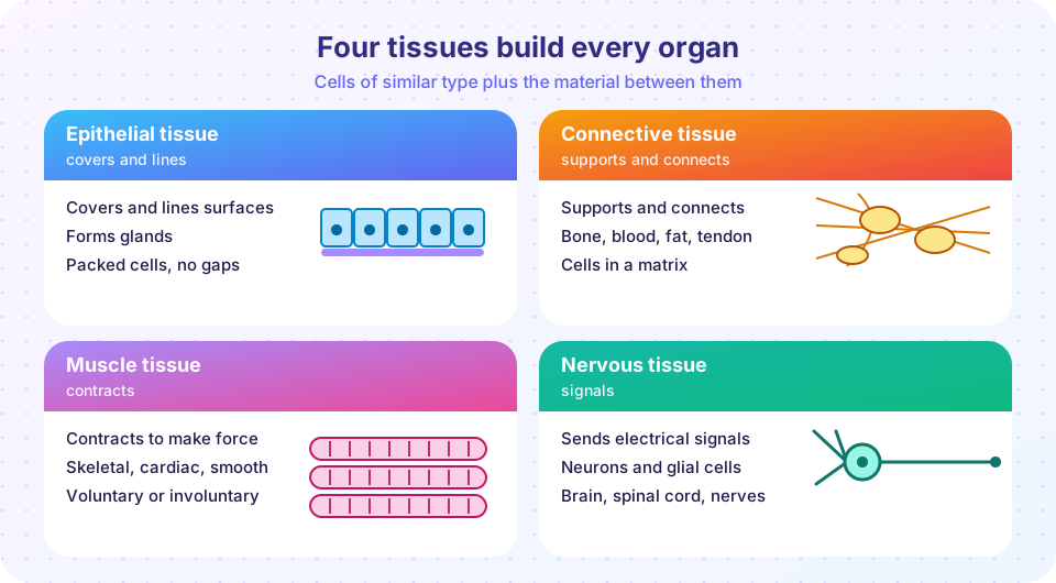
What you will learn
- The location, structure and function of the four tissue types.
- How epithelium is named by cell shape and layering.
- The main kinds of connective tissue and muscle tissue.
- How tissues repair themselves and how body membranes are built.
- The layers of the skin.
Key terms
- Matrix: the non-living material between cells in a tissue, made of fibers and ground substance.
- Epithelium: tissue that covers surfaces and lines cavities and forms glands.
- Basement membrane: a thin supporting layer that anchors epithelium to the tissue below.
- Avascular: having no blood vessels of its own.
- Neuroglia (glial cells): supporting cells of nervous tissue.
Epithelial tissue
Epithelial tissue forms the covering of the body (the outer skin), the lining of hollow organs and body cavities, and glands. It has these features:
- Cells are packed closely together with very little matrix, forming continuous sheets.
- One side is free, facing the outside world or an interior space (the apical surface); the other side rests on a basement membrane.
- It is avascular: nutrients arrive by diffusion from the connective tissue below. It has nerve endings, though.
- It regenerates quickly: skin cells and the cells lining the intestine are replaced in days to weeks.
Epithelium is named by combining two descriptions. The first word describes the number of layers: simple (one layer, for absorption, secretion and filtration) or stratified (several layers, for protection). The second describes the shape of the cells at the free surface: squamous (flat and scale-like), cuboidal (cube-like) or columnar (tall and column-like).
| Type | Where it is found | Main job |
|---|---|---|
| Simple squamous | air sacs of the lungs, capillary walls, lining of blood vessels | quick diffusion and filtration across a thin wall |
| Simple cuboidal | kidney tubules, small glands | secretion and absorption |
| Simple columnar | stomach and intestinal lining | absorption and secretion; goblet cells make mucus |
| Pseudostratified ciliated columnar | trachea and airways | one layer that looks layered; cilia sweep mucus |
| Stratified squamous | skin surface, mouth, esophagus, vagina | protection from abrasion |
| Transitional | bladder and ureters | stretches without tearing |
The surface layer of the skin is stratified squamous epithelium whose top cells are filled with the tough protein keratin and are dead; in the mouth and esophagus the same pattern appears without keratin and stays moist.
Glands are made of epithelial cells that secrete. Exocrine glands release products through ducts onto a surface or into a cavity: sweat, oil, saliva, mucus, digestive enzymes. Endocrine glands have no ducts and release hormones into the blood (Chapter 5).
Connective tissue
Connective tissue is the most abundant and varied tissue. It binds, supports, protects, insulates and transports. Unlike epithelium, it has relatively few cells scattered in a large matrix, made of protein fibers and ground substance. The three fiber types are collagen (strong and flexible, the most abundant protein in the body), elastic fibers (stretch and recoil) and reticular fibers (fine supporting mesh). The ground substance can be fluid, gel-like or solid.
| Connective tissue | Matrix and cells | Typical locations | Function |
|---|---|---|---|
| Loose (areolar) | gel with scattered fibers | under skin and epithelia, around organs | cushions, holds fluid, supports |
| Adipose | cells packed with fat droplets | under the skin, around kidneys and heart | energy storage, insulation, padding |
| Dense regular | parallel collagen fibers | tendons, ligaments | strong pull in one direction |
| Dense irregular | collagen fibers in all directions | dermis of the skin, organ capsules | strength in many directions |
| Hyaline cartilage | firm, glassy matrix with chondrocytes | nose, rib ends, joint surfaces | smooth support, cushions |
| Bone | hard matrix of calcium salts and collagen, with osteocytes | skeleton | support, protection, mineral storage |
| Blood | liquid matrix (plasma) with red cells, white cells and platelets | inside blood vessels | transport and defense |
Cartilage has no blood vessels (it is avascular), which is why damaged cartilage heals slowly, and tendons, with a poor blood supply, also heal slowly. Bone is well supplied with blood and heals much better. Blood is unusual because its matrix is a liquid, and Chapter 5 covers it in depth.
Muscle tissue
Muscle tissue is specialized to contract, shortening to produce force. There are three kinds.
- Skeletal muscle is attached to bones. Its long cylindrical cells, called fibers, are multinucleate and striated (striped, from the regular arrangement of contractile proteins). It is under voluntary control.
- Cardiac muscle forms the wall of the heart. Cells are striated but branched, usually with a single nucleus, joined end to end by intercalated discs that pass the signal to contract from cell to cell. It is involuntary and rhythmic.
- Smooth muscle forms the walls of hollow organs and blood vessels. Its spindle-shaped cells have one nucleus and are not striated. It is involuntary and slower, and it moves food along the gut and adjusts blood vessel width.
You will study how muscle contracts in Chapter 3.
Nervous tissue
Nervous tissue carries information as electrical signals. It consists of neurons, cells with a cell body, branching dendrites that receive signals and an axon that sends them, and neuroglia, which support, insulate and nourish neurons. It makes up the brain, spinal cord and nerves, and it is the subject of Chapter 4.
Tissue repair
When tissue is damaged, the body can respond by regeneration, replacing the lost cells with the same type of cells (skin, intestinal lining, bone and liver do this well), or by fibrosis, filling the gap with dense connective tissue, called scar tissue, made mostly of collagen (this is typical for heart muscle, which regenerates poorly, and for deep skin wounds). A scar holds the tissue together but cannot do the job of the original tissue. Healing has overlapping stages: bleeding stops (clotting), inflammation clears debris, new tissue forms and the area is remodeled over weeks to months. Good nutrition, blood supply and a clean wound speed healing.
Body membranes
Tissues also combine into thin sheets that cover and line:
- The cutaneous membrane is the skin (epithelium on connective tissue).
- Mucous membranes line passages open to the outside, such as the mouth, digestive tract and airways. Their epithelium sits on loose connective tissue and is often moistened by mucus.
- Serous membranes line closed cavities (pleura, pericardium, peritoneum, covered in Chapter 1).
- Synovial membranes are connective tissue only and line the cavities of freely movable joints, secreting lubricating synovial fluid.
The skin
The skin (integumentary system) is a membrane and an organ in one. In adults it covers 1.5 to 2 square meters and weighs a few kilograms. It has three layers.
- Epidermis, the outer layer, is keratinized stratified squamous epithelium. New cells are made in the deepest layer (the stratum basale), push upward over about a month as they fill with keratin and die, and are shed from the top. The epidermis also contains melanocytes, which make the pigment melanin to absorb ultraviolet light and protect DNA, plus touch receptors and immune cells. The epidermis has no blood vessels.
- Dermis, below it, is dense irregular connective tissue with collagen and elastic fibers, blood vessels, nerve endings, hair follicles, and sweat and oil glands. It gives the skin strength and stretch.
- Hypodermis (subcutaneous tissue), under the dermis, is mostly loose and fatty tissue that anchors the skin, insulates and stores energy. Strictly it is not part of the skin.
Skin protects against germs, water loss and sunlight; helps regulate temperature through sweating and blood flow; senses touch, pressure, temperature and pain; excretes small amounts of salts and wastes in sweat; and makes vitamin D when exposed to ultraviolet light. Burns are classified by depth: a first-degree burn affects only the epidermis, a second-degree (partial thickness) burn reaches into the dermis, and a third-degree (full thickness) burn destroys the full depth of skin.
Classify the tissue
Histologists identify epithelium by two questions: how many layers, and what shape are the surface cells? The program below encodes that as a small decision function and runs it on several examples. It is a teaching model of the naming rules, not a diagnostic tool.
Press Run (or Ctrl + Enter) to see the result here.
Program input · what you'd type while the program runs
Type the answers before you press Run, one per line. Each time the program reads a line (with input()), it gets the next line from this box.
Common misconceptions
- "Skin is only a covering." It is an organ with sensing, temperature control, defense and vitamin D production.
- "Epithelial tissue only covers the outside." It also lines the airways, gut, blood vessels and forms glands.
- "Bone is dead, like a rock." Bone is living tissue with blood vessels, cells and constant remodeling.
- "Connective tissue just holds things together." It also stores fat, carries blood and cushions joints.
- "Cardiac muscle regenerates like skin." Heart muscle repairs mostly by scarring, which is why damage after a heart attack is long-lasting.
Check your understanding
Quick check · 5 questions
of 5 answered You scored / 5 · perfect!
-
1. A tissue made of one layer of flat cells is best suited to which job?
Simple squamous epithelium is thin, so substances cross it quickly, as in air sacs and capillary walls. -
2. Which tissue forms tendons?
Tendons are dense regular connective tissue with parallel collagen fibers that resist pulling in one direction. -
3. Which statement about cardiac muscle is correct?
Cardiac muscle is striated and involuntary and its cells link through intercalated discs. -
4. Which layer of the skin contains blood vessels, nerves and glands?
The dermis is the vascular, nerve-rich connective layer under the epidermis. -
5. Why does cartilage heal slowly?
Cartilage is avascular, so nutrients and repair cells arrive slowly by diffusion.
Summary
- The four primary tissues are epithelial, connective, muscle and nervous.
- Epithelium is named by layers (simple or stratified) and surface cell shape (squamous, cuboidal or columnar), and it lines or covers surfaces and forms glands.
- Connective tissue has few cells in a large matrix and includes loose, adipose, dense, cartilage, bone and blood.
- Muscle tissue is skeletal, cardiac or smooth; nervous tissue is neurons plus neuroglia.
- Tissues repair by regeneration or by scarring; body membranes are cutaneous, mucous, serous or synovial.
- The skin has an epidermis, a dermis and an underlying hypodermis.
Next: Chapter 2 review
Track your progress
Create a free account to tick off lessons and pick up where you left off on any device.
of Anatomy and Physiology lessons completed