


















Lesson 27 of 52 · Endocrine and Cardiovascular Systems
The heart: chambers, valves and the cardiac cycle
Learn the four chambers and valves of the heart, trace blood through both circuits, and read the cardiac cycle, heart sounds and a basic ECG.
Your heart is a fist-sized muscle that beats about 100,000 times a day without a rest. It pumps roughly five liters of blood every minute at rest, and several times that during hard exercise. For centuries people thought blood was made in the liver and used up by the body. In 1628 the English physician William Harvey published a short book arguing from careful measurement that the heart pumps blood in a circle. He estimated that the heart ejects far more blood in an hour than the body weighs, so the blood must be returning, not being made anew. He lacked a microscope and could not see capillaries, which Marcello Malpighi observed a few decades later and which completed Harvey's picture.
This lesson covers the heart's structure, the path of blood through it, the timing of one heartbeat, the electrical system that drives it, and how an electrocardiogram records it.
What you will learn
- Name the four chambers, four valves and main vessels of the heart.
- Trace a red blood cell through the pulmonary and systemic circuits.
- Describe the phases of the cardiac cycle and what causes the heart sounds.
- Explain how the sinoatrial node starts a heartbeat and how the signal spreads.
- Calculate cardiac output from heart rate and stroke volume.
Key terms
Atrium: an upper chamber that receives blood returning to the heart.
Ventricle: a lower chamber that pumps blood out of the heart.
Cardiac output: the volume of blood pumped by each ventricle per minute.
Systole and diastole: the contraction and relaxation phases of the cardiac cycle.
ECG: a recording of the heart's electrical activity from the body surface.
Structure: a double pump
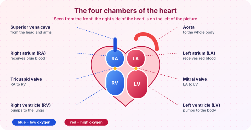
The heart is really two pumps side by side, separated by a muscular wall called the septum. The right side receives blood that has given up its oxygen and sends it to the lungs. The left side receives blood freshly loaded with oxygen and sends it to the rest of the body.
Each side has two chambers. The atria (singular atrium) are thin-walled receiving rooms. The ventricles are thick-walled pumps. The left ventricle has the thickest wall, about three times as thick as the right, because it must push blood through the long systemic circuit at higher pressure, while the right ventricle only pushes blood through the nearby lungs.
Four valves make flow one-way. Between each atrium and ventricle sits an atrioventricular (AV) valve: the tricuspid valve on the right (three flaps) and the mitral or bicuspid valve on the left (two flaps). Thin cords called chordae tendineae anchor the flaps to small muscles in the ventricle so they do not blow back into the atrium. At the exit of each ventricle sits a semilunar valve with three half-moon cups: the pulmonary valve (to the pulmonary artery) and the aortic valve (to the aorta).
The heart wall has three layers: the thin inner endocardium, the thick muscle myocardium and the outer epicardium, all inside a fibrous sac, the pericardium. The heart muscle itself is fed by the coronary arteries, the first branches of the aorta. A blockage in one of these is what causes a heart attack.
The path of blood
Follow one red blood cell around the double circuit:
- Blood low in oxygen returns from the body by the superior and inferior vena cava into the right atrium.
- It passes the tricuspid valve into the right ventricle.
- The right ventricle pumps it through the pulmonary valve into the pulmonary arteries, the only arteries that carry low-oxygen blood, to the lungs. This is the pulmonary circuit.
- In lung capillaries the blood picks up oxygen and loses carbon dioxide.
- Pulmonary veins, the only veins that carry high-oxygen blood, bring it to the left atrium.
- It passes the mitral valve into the left ventricle.
- The left ventricle pumps it through the aortic valve into the aorta and the systemic circuit to every tissue.
Both circuits are in series, so the same volume must pass through each every minute. If the left side pumped even slightly more than the right, blood would pile up in the lungs.
The cardiac cycle
One heartbeat is a sequence of contraction (systole) and relaxation (diastole). At a resting rate of 75 beats per minute one cycle lasts 0.8 seconds.
- Late diastole. All chambers relax. Blood flows from the veins through the atria and ventricles. About 70 percent of ventricular filling happens passively.
- Atrial systole. The atria contract and push the last 20 to 30 percent of blood into the ventricles.
- Isovolumetric contraction. The ventricles begin to contract. Pressure rises and shuts the AV valves, but is not yet high enough to open the semilunar valves, so volume is constant for a moment.
- Ventricular ejection. Ventricular pressure exceeds the pressure in the aorta and pulmonary artery. The semilunar valves open and blood is ejected.
- Isovolumetric relaxation. The ventricles relax, pressure falls, the semilunar valves snap shut, and the cycle repeats.
The two normal heart sounds are caused by valve closure. The first ("lub") is the closing of the AV valves at the start of ventricular systole; the second ("dub") is the closing of the semilunar valves at the start of diastole. A murmur is an extra sound often caused by turbulent flow, for example through a narrowed or leaking valve. Not every murmur is a sign of disease, but a clinician must interpret it.
The volume of blood ejected by one ventricle in one beat is the stroke volume (about 70 mL at rest in an adult). Multiply by heart rate to get cardiac output:
cardiac output = heart rate × stroke volume
At 72 beats per minute and 70 mL per beat this is about 5,040 mL, or roughly 5 L/min. In hard exercise an average person can raise it to 20 L/min or more; elite endurance athletes can reach higher values. The heart can increase its stroke volume because stretched cardiac muscle contracts more forcefully, a relationship described by Otto Frank in the 1890s and Ernest Starling in the early 1900s (the Frank-Starling law).
The electrical system
Cardiac muscle cells are linked end to end by intercalated discs containing gap junctions, tiny channels that let electrical current pass from cell to cell. A wave of depolarization therefore spreads through the heart almost like a single unit. Even more remarkably, the heart generates its own rhythm: a heart removed from the body continues to beat for a while.
The pacemaker is a cluster of specialized cells in the right atrium, the sinoatrial (SA) node. Its membrane slowly depolarizes by itself until it fires, usually 60 to 100 times a minute. From there:
- The wave spreads across both atria, causing them to contract.
- It reaches the atrioventricular (AV) node, where it is held for about 0.1 second. This delay lets the atria finish emptying into the ventricles.
- It then travels down the bundle of His and its left and right branches along the septum.
- Purkinje fibers spread it quickly through the ventricle walls so they contract from the apex upward, squeezing blood toward the exit valves.
The nervous system and hormones adjust the rate. The sympathetic nerves and adrenaline speed the SA node and strengthen contraction; the vagus nerve (parasympathetic) slows it. This is why your heart rate rises when you stand up quickly and falls during sleep.
The ECG
In 1903 Willem Einthoven developed a sensitive string galvanometer that recorded the heart's electrical signals from the body surface; he received the 1924 Nobel Prize for it. A modern electrocardiogram (ECG) has the same three main waves.
| Wave | Represents |
|---|---|
| P wave | Depolarization of the atria |
| QRS complex | Depolarization of the ventricles (the atria repolarize during this time but their signal is hidden) |
| T wave | Repolarization of the ventricles |
The PR interval reflects the AV node delay and the ST segment the plateau phase of ventricular cells. Changes in these shapes help clinicians detect rhythm problems and heart attacks. An ECG records electrical activity, not pumping, so a heart can show electrical activity without effective pumping.
Common misconceptions
"The left side of the heart is on the left of the picture." In standard diagrams the patient's left is on the viewer's right, as if the patient faces you.
"Arteries carry red blood and veins carry blue blood." Arteries carry blood away from the heart; veins carry it toward the heart. Pulmonary arteries carry low-oxygen blood. Real veins look blue through skin, but blood in veins is dark red, not blue.
"The heart beats because the brain tells each beat." The brain only adjusts the rate; the SA node sets each beat.
Try it
The first TryIt draws a stylized ECG from the sum of five Gaussian bumps. It is a schematic shape, not real patient data.
Press Run (or Ctrl + Enter) to see the result here.
Program input · what you'd type while the program runs
Type the answers before you press Run, one per line. Each time the program reads a line (with input()), it gets the next line from this box.
The second TryIt calculates cardiac output and animates the heart at the chosen rate. It is a simple model of the formula, not of the full physiology.
Check your understanding
Quick check · 6 questions
of 6 answered You scored / 6 · perfect!
-
1. Which valve lies between the left atrium and the left ventricle?
The mitral or bicuspid valve separates the left atrium from the left ventricle. The tricuspid valve is on the right. -
2. Why is the wall of the left ventricle thicker than the wall of the right ventricle?
The systemic circuit has high resistance, so the left ventricle needs more muscle than the right, which only supplies the lungs. -
3. What causes the first heart sound, 'lub'?
The first sound marks the closure of the tricuspid and mitral valves at the start of ventricular systole. -
4. What is the purpose of the delay at the AV node?
The roughly 0.1 s delay coordinates atrial and ventricular contraction so the ventricles are full before they pump. -
5. A person has a heart rate of 60 beats per minute and a stroke volume of 80 mL. What is the cardiac output?
60 × 80 mL = 4,800 mL, which is 4.8 L per minute. -
6. Which ECG wave shows ventricular depolarization?
The QRS complex is the large deflection produced when the ventricles depolarize.
Summary
- The heart is two pumps in series: the right side serves the lungs, the left side serves the body.
- Four valves keep flow one-way; their closing makes the heart sounds.
- The cardiac cycle alternates systole and diastole; cardiac output equals heart rate times stroke volume.
- The SA node sets the rhythm; the AV node delays the signal; Purkinje fibers spread it through the ventricles.
- The ECG records P, QRS and T waves for atrial depolarization, ventricular depolarization and ventricular repolarization.
Track your progress
Create a free account to tick off lessons and pick up where you left off on any device.
of Anatomy and Physiology lessons completed