


















Lesson 23 of 52 · Nervous System and Senses
Chapter 4 review: the nervous system and senses
Review neurons, brain regions, the spinal cord, reflexes, the autonomic system and the senses with a recap table, pathway tracer and ten-question quiz.
Chapter 4 followed information from the world to the brain and back out to the muscles. You saw how a neuron builds a voltage spike from ion gradients, how synapses pass it on, how the brain and spinal cord process it, how nerves and the autonomic system carry commands, and how the special senses turn light, sound and chemicals into signals. This lesson connects those pieces into whole stories and tests you on them.
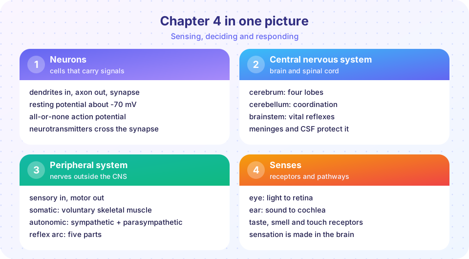
What you will learn
- A one-page recap of the chapter.
- How to trace a signal through several structures in the right order.
- How to apply several lessons to one situation.
Recap table
| Topic | Remember |
|---|---|
| Organization | CNS (brain, spinal cord) and PNS; sensory (afferent) and motor (efferent); somatic and autonomic |
| Cells | neurons carry signals (about 86 billion); glia support, insulate and protect |
| Resting potential | about -70 mV from ion gradients, K⁺ leak channels and the Na⁺/K⁺ pump |
| Action potential | threshold about -55 mV; Na⁺ in, then K⁺ out; all-or-none; frequency coding |
| Conduction | myelin and nodes of Ranvier give saltatory conduction (up to about 100 m/s) |
| Synapse | Ca²⁺ triggers vesicle release; neurotransmitter binds receptors; then cleared |
| Protection | skull, meninges (dura, arachnoid, pia), cerebrospinal fluid, blood-brain barrier |
| Brain | frontal, parietal, temporal and occipital lobes; thalamus, hypothalamus, brainstem, cerebellum |
| Spinal cord | gray matter horns, white matter tracts, 31 pairs of spinal nerves, reflexes |
| Reflex arc | receptor, sensory neuron, integration center, motor neuron, effector |
| Autonomic | sympathetic (thoracolumbar, norepinephrine) and parasympathetic (craniosacral, ACh); enteric network |
| Senses | rods and cones, lens and cornea; ossicles, cochlea and hair cells; vestibular organs; taste and smell receptors |
A worked example: hearing your name
Someone calls your name across a room and you turn your head. Many parts of the chapter work in sequence.
- Receptor and transduction. Sound waves vibrate the eardrum, the ossicles amplify the vibration and the stapes pushes on the oval window. Waves in the cochlear fluid bend hair cells, which release neurotransmitter.
- Afferent pathway. The cochlear branch of cranial nerve VIII carries action potentials to the brainstem, then to the thalamus and then to the primary auditory cortex in the temporal lobe.
- Integration. Temporal lobe regions, including Wernicke's area, recognize the sounds as language and as your name. The frontal lobe decides to respond.
- Efferent pathway. The primary motor cortex sends commands down the corticospinal tract, which crosses to the other side. Spinal motor neurons in the cervical cord activate neck muscles at neuromuscular junctions using acetylcholine.
- Fine control. The cerebellum smooths the movement, and proprioceptors in the neck report the head's position.
- Autonomic echo. If the voice sounds urgent, the sympathetic system may raise your heart rate slightly.
The whole sequence takes a few tenths of a second. Notice how the same ideas return: transduction, synapses, tracts, integration and effectors.
Trace the pathway
The program below stores several pathways as ordered lists of structures and prints them step by step. It is a teaching model that reflects the textbook sequences from this chapter, and it is easy to add your own pathways.
Press Run (or Ctrl + Enter) to see the result here.
Program input · what you'd type while the program runs
Type the answers before you press Run, one per line. Each time the program reads a line (with input()), it gets the next line from this box.
Test your recall with the chapter deck. Answer aloud before revealing.
Where students trip up
Three habits cost marks. First, confusing afferent and efferent: remember that afferent signals arrive at the CNS and efferent signals exit. Second, mixing up the two autonomic divisions' structure: sympathetic has short preganglionic fibers (ganglia near the cord), parasympathetic has long preganglionic fibers (ganglia near the organ). Third, treating action potentials as variable in size: they are all-or-none, and intensity is carried by frequency. If you can draw the action potential graph and label each phase and ion movement from memory, you have most of the chapter.
Common misconceptions
- "Reflexes need the brain." Spinal reflexes are processed in the cord. The brain finds out afterward.
- "Sympathetic and parasympathetic never act together." Both operate continuously, and the balance shifts.
- "A stronger stimulus gives a bigger action potential." Spikes are all-or-none; strength changes the rate.
Check your understanding
Quick check · 10 questions
of 10 answered You scored / 10 · perfect!
-
1. Which event begins the depolarization of an action potential?
Sodium entry through voltage-gated channels drives the rising phase. -
2. What does the thalamus do?
The thalamus is the main sensory relay of the brain. -
3. Which neurotransmitter is released at the neuromuscular junction?
Motor neurons release acetylcholine onto skeletal muscle fibers. -
4. Where does the primary visual cortex lie?
Visual information is processed first in the occipital lobe. -
5. Which is a parasympathetic effect?
Parasympathetic activity promotes digestion, slows the heart and constricts the pupils. -
6. Which structure produces cerebrospinal fluid?
Choroid plexuses make CSF at roughly 500 mL per day. -
7. The knee-jerk reflex is monosynaptic. What does that mean?
The sensory neuron synapses directly on the motor neuron, so there is a single central synapse. -
8. What converts sound vibrations into nerve signals?
Hair cells on the basilar membrane in the cochlea transduce vibration into electrical signals. -
9. Which cells make up most of the cells in the retina's central pit for sharp color vision?
The fovea is packed with cones. -
10. How does the nervous system code a stronger stimulus?
Action potentials are all-or-none, so intensity is coded by frequency and recruitment.
Summary
- Neurons use ion gradients to build the resting potential and all-or-none action potentials; synapses pass signals chemically.
- The CNS is protected by bone, meninges, CSF and the blood-brain barrier; the brain's regions specialize, and the spinal cord carries tracts and runs reflexes.
- The PNS has 12 cranial and 31 spinal nerve pairs; reflex arcs have five parts; the autonomic system uses two-neuron pathways in sympathetic and parasympathetic divisions.
- The senses transduce light, sound, chemicals and mechanical stimuli into signals that the brain turns into perception.
Next: Chapter 5 overview: Endocrine and Cardiovascular Systems
Track your progress
Create a free account to tick off lessons and pick up where you left off on any device.
of Anatomy and Physiology lessons completed