


















Lesson 3 of 52 · Getting Started
Body cavities, membranes and abdominal regions
Map the dorsal and ventral body cavities, the pleura, pericardium and peritoneum, and the four quadrants and nine regions used to locate organs and pain.
Your organs are not floating loose. Each sits in a protective space called a body cavity, lined with slippery membranes that let organs move without rubbing themselves raw. Your heart beats about 100,000 times a day, your lungs inflate and deflate about 20,000 times, and your intestines squirm constantly, yet none of this wears anything out because the cavities and their membranes handle the friction.
Knowing which cavity holds which organ is the foundation for understanding symptoms, scans and surgery. A doctor who hears "sharp pain in the right lower quadrant" immediately thinks of a short list of organs. This lesson gives you the map.
What you will learn
- The two main body cavities and their subdivisions.
- How serous membranes (pleura, pericardium, peritoneum) protect organs.
- Which organs sit in which cavity.
- How the four quadrants and nine regions are used to locate organs and pain.
Key terms
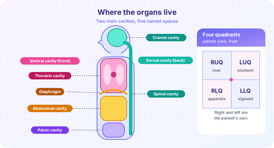
- Dorsal body cavity: the cranial cavity (brain) plus the spinal cavity (spinal cord).
- Ventral body cavity: the thoracic cavity plus the abdominopelvic cavity.
- Serous membrane: a thin, two-layer membrane that secretes a small amount of lubricating fluid.
- Parietal / visceral layer: the layer lining the cavity wall / the layer covering the organ.
- Mediastinum: the central part of the thoracic cavity between the lungs, holding the heart and great vessels.
The dorsal cavity: the control center
The dorsal body cavity runs along the back of the body. It has two continuous parts. The cranial cavity, inside the skull, houses the brain. The spinal (vertebral) cavity, the canal formed by the stacked vertebrae, houses the spinal cord. Both are protected by bone, and both are wrapped in three layers of membrane called the meninges. The brain and spinal cord also float in cerebrospinal fluid, which cushions them. In Chapter 4 you will see how this protects the most delicate tissue in the body.
The ventral cavity: the working organs
The ventral body cavity is much larger and sits at the front. It is split by the diaphragm, a dome-shaped sheet of skeletal muscle and the main muscle of breathing, into two parts.
Thoracic cavity
The thoracic cavity is surrounded by the rib cage. It contains three smaller spaces:
- Two pleural cavities, one around each lung.
- The pericardial cavity, a small space around the heart.
- The mediastinum, the central region between the lungs. It holds the heart, the thymus, the trachea (windpipe), the esophagus (food tube) and the large blood vessels entering and leaving the heart. The lungs themselves are not in the mediastinum.
Abdominopelvic cavity
Below the diaphragm lies the abdominopelvic cavity. No wall separates its two halves, but they are named for their position:
- The abdominal cavity holds the stomach, liver, gallbladder, spleen, pancreas, small intestine and most of the large intestine. The kidneys sit against the back wall of this region.
- The pelvic cavity, inside the bony pelvis, holds the urinary bladder, the rectum and, depending on sex, the internal reproductive organs.
Small cavities elsewhere
You also have several small cavities that are not part of the two main ones: the oral cavity (mouth), the nasal cavity, the orbital cavities (eye sockets), the middle ear cavities and the synovial cavities inside freely movable joints.
Serous membranes: built-in lubrication
Think of pushing your fist into a partly inflated balloon. The balloon wraps around your fist in two layers: one touching your fist and one forming the outer surface. A serous membrane is arranged the same way. The visceral layer (from the Latin viscera, internal organs) covers the surface of the organ. The parietal layer (from paries, wall) lines the cavity wall. Between them is a thin gap, the serous cavity, filled with a few milliliters of serous fluid, a watery lubricant that lets the two layers glide over each other.
| Membrane | Where | Surrounds |
|---|---|---|
| Pleura | thoracic cavity | each lung |
| Pericardium | mediastinum | the heart |
| Peritoneum | abdominopelvic cavity | abdominal organs |
Infection or irritation of these membranes causes pleurisy, pericarditis and peritonitis. (The ending -itis means inflammation.) They are painful because the two layers, normally slick, begin to rub.
Not every abdominal organ is wrapped by the peritoneum. Organs that lie behind it, against the back wall, are retroperitoneal. The kidneys, adrenal glands and most of the pancreas are examples. Organs hanging inside the peritoneal cavity are held in place by double-layered folds called mesenteries, which also carry blood vessels and nerves to the intestines.
Locating things: quadrants and regions
The abdominopelvic cavity is large, so clinicians divide it into zones. The simplest method uses two lines that cross at the navel: a vertical midline and a horizontal line, giving four quadrants.
| Quadrant | Abbreviation | Main organs |
|---|---|---|
| Right upper | RUQ | most of the liver, gallbladder, part of the large intestine |
| Left upper | LUQ | stomach, spleen, part of the pancreas |
| Right lower | RLQ | appendix, part of the small and large intestine |
| Left lower | LLQ | sigmoid colon, part of the small intestine |
Remember that "right" and "left" are the patient's sides. Doctors use the quadrants for quick descriptions. Pain starting around the navel and then settling in the RLQ is a classic pattern in appendicitis, which is why the RLQ gets special attention in emergency rooms.
For more precision, anatomists use nine regions formed by two vertical and two horizontal lines, like a tic-tac-toe grid:
| Right | Middle | Left | |
|---|---|---|---|
| Upper | right hypochondriac | epigastric | left hypochondriac |
| Middle | right lumbar | umbilical | left lumbar |
| Lower | right iliac (inguinal) | hypogastric (pubic) | left iliac (inguinal) |
The word epigastric means "upon the stomach," and hypogastric means "below the stomach," which is a good way to remember that they sit above and below the umbilical region.
Practice with a lookup table
The next demonstration stores the cavity, quadrant and membrane for a set of organs as a dictionary. It is a teaching model of the facts above: real organs are bigger than a single quadrant, so we list the quadrant where most of the organ lies.
Press Run (or Ctrl + Enter) to see the result here.
Program input · what you'd type while the program runs
Type the answers before you press Run, one per line. Each time the program reads a line (with input()), it gets the next line from this box.
Common misconceptions
- "The lungs are in the mediastinum." The mediastinum is the space between the lungs. Each lung has its own pleural cavity.
- "The abdominal and pelvic cavities are separate rooms." They are one continuous space, the abdominopelvic cavity, named in two parts for convenience.
- "The serous cavity is full of fluid." It holds only a thin film, a few milliliters, just enough to lubricate. A true buildup (an effusion) is a sign of a problem.
- "The liver is only on the right." Most of it is in the right upper quadrant, but a lobe extends across the midline toward the left.
Check your understanding
Quick check · 5 questions
of 5 answered You scored / 5 · perfect!
-
1. Which two cavities together make up the dorsal body cavity?
The cranial cavity (brain) and spinal cavity (spinal cord) are continuous and form the dorsal cavity. -
2. What is the role of serous fluid?
Serous fluid fills the thin space between the visceral and parietal layers and reduces friction. -
3. Which organ is retroperitoneal?
The kidneys lie behind the peritoneum against the back wall of the abdomen. -
4. A doctor describes pain in the LUQ. Which organ is a likely source?
The stomach and spleen lie mainly in the left upper quadrant. The appendix is in the RLQ and the gallbladder in the RUQ. -
5. The epigastric region lies in which part of the nine-region grid?
Epigastric means upon the stomach: it is the upper middle region.
Summary
- The dorsal cavity (cranial and spinal) holds the brain and spinal cord; the ventral cavity holds most other organs.
- The diaphragm separates the thoracic cavity from the abdominopelvic cavity.
- The thoracic cavity has two pleural cavities, the pericardial cavity and the mediastinum.
- Serous membranes have a visceral and a parietal layer with a thin film of fluid between them.
- Four quadrants (RUQ, LUQ, RLQ, LLQ) and nine regions help locate organs and pain.
Next: The eleven organ systems
Track your progress
Create a free account to tick off lessons and pick up where you left off on any device.
of Anatomy and Physiology lessons completed