


















Lesson 22 of 52 · Nervous System and Senses
The senses: touch, vision, hearing, balance, taste and smell
See how receptors work and how the eye, ear, tongue and nose turn light, sound and chemicals into nerve signals, plus touch, pain and balance.
Everything you know about the world reaches you through sensory receptors, specialized cells or nerve endings that convert one kind of energy (light, sound waves, pressure, chemicals, heat) into the electrical language of the nervous system. This conversion is called transduction. Your brain never touches a red apple or hears a violin directly; it receives patterns of action potentials from receptors and builds the experience of color or music out of them. In that sense, sensation is made in the brain.
This lesson starts with the general principles of receptors and the senses of touch and pain, then follows the light entering the eye, the sound entering the ear, and the chemicals that you smell and taste.
What you will learn
- How receptors are classified and what sensory adaptation is.
- The receptors for touch, pressure, temperature, pain and body position.
- The structure of the eye, how it focuses and how the retina detects light.
- The structure of the ear and how hearing and balance work.
- How taste and smell detect chemicals.
Key terms
- Receptor: a sensory structure that detects a stimulus and starts a nerve signal.
- Transduction: conversion of a stimulus into an electrical signal in a receptor.
- Adaptation: a decrease in response to a constant stimulus over time.
- Photoreceptor: a retinal cell (rod or cone) that detects light.
- Cochlea: the coiled, fluid-filled part of the inner ear that detects sound.
Receptors and the general senses
Receptors are classed by the stimulus they detect: mechanoreceptors (touch, pressure, stretch, vibration, sound), thermoreceptors (temperature), nociceptors (pain), chemoreceptors (taste, smell, blood chemistry) and photoreceptors (light). The general senses are spread over the body, and the special senses (vision, hearing, balance, taste and smell) belong to dedicated sense organs in the head.
Most receptors show adaptation. If you put on a shirt, you feel it at first and then stop noticing it, because touch receptors fire mainly when something changes. Adaptation spares the brain from processing unchanging background information. Pain receptors adapt very little, which suits their warning role.
Touch, temperature, pain and position
The skin holds several kinds of mechanoreceptor. Meissner corpuscles in the upper dermis, dense in fingertips, detect light touch. Merkel discs respond to sustained light pressure and fine texture. Pacinian corpuscles deep in the skin are onion-like structures that detect vibration and deep pressure. Ruffini endings sense stretch. Free nerve endings, simple bare branches, detect temperature, pain and itch. Fingertips and lips have many receptors, so two close points feel like two; on the back they feel like one.
Pain is carried by nociceptors that respond to damaging stimuli, mainly through thin fibers (fast, sharp pain via Aδ fibers; slow, dull pain via C fibers, as you saw in the overview). Pain from internal organs is often felt in the skin, called referred pain. Heart pain, for example, may be felt in the left shoulder and arm, because signals from the heart and from that skin area converge on the same spinal cord neurons. Proprioceptors such as muscle spindles and Golgi tendon organs report muscle length and tension so that you know where your limbs are without looking.
Vision
Structure of the eye
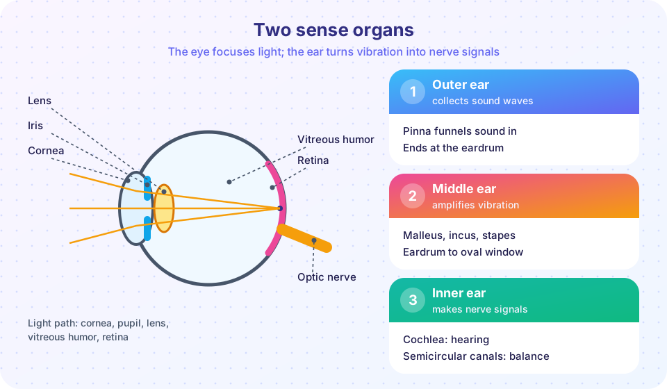
The eye is a sphere about 2.4 centimeters across with three layers. The outer fibrous layer consists of the white sclera and the transparent cornea at the front, which does most of the focusing (about two thirds of the eye's optical power). The middle vascular layer includes the choroid (blood supply and dark pigment that absorbs stray light), the ciliary body (which holds and reshapes the lens and makes aqueous humor) and the iris, the colored ring that surrounds the pupil. The inner layer is the retina, the light-sensitive neural tissue. The lens sits behind the iris. The chamber in front of the lens is filled with watery aqueous humor, and the large chamber behind it with the clear gel called vitreous humor.
Light takes this path: cornea, aqueous humor, pupil, lens, vitreous humor, retina. The iris adjusts the pupil: in bright light the pupil constricts (parasympathetic), and in dim light it dilates (sympathetic).
Focusing
The cornea and lens bend (refract) light to form a small, sharp, upside-down image on the retina; the brain interprets it the right way up. The cornea has fixed power, and the lens changes it to focus at different distances. This adjustment is called accommodation. To look at a near object, the ciliary muscle contracts, which relaxes the fibers that hold the lens, and the lens rounds up and becomes more powerful. For a far object, the muscle relaxes, the fibers tighten and the lens flattens. With age the lens stiffens and loses its ability to change shape, so that near objects become blurry from the mid-40s; this is presbyopia.
Common refractive errors arise when the eyeball and the optics do not match. In myopia (nearsightedness), the eyeball is too long for its optics or the optics are too strong, so distant objects focus in front of the retina and look blurred. In hyperopia (farsightedness), the opposite is true. Astigmatism is caused by uneven curvature of the cornea. Glasses or contact lenses correct these errors.
Press Run (or Ctrl + Enter) to see the result here.
Program input · what you'd type while the program runs
Type the answers before you press Run, one per line. Each time the program reads a line (with input()), it gets the next line from this box.
The retina
The retina has layers of neurons. Light passes through them to reach the photoreceptors at the back, which are the rods and the cones. The signal then moves forward through bipolar cells to ganglion cells, whose axons form the optic nerve.
- Rods (about 120 million) are very sensitive and work in dim light, but give only black-and-white, low-detail vision. They are mostly in the periphery.
- Cones (about 6 million) work in bright light, give sharp color vision and are concentrated in the fovea, a small pit in the center of the retina where vision is sharpest. There are three kinds of cones, which respond best to short (blue), medium (green) and long (red) wavelengths. The brain compares their outputs to create color.
In the dark, photoreceptors are depolarized and release glutamate steadily. Light makes a pigment molecule (rhodopsin in rods, which contains a form of vitamin A) change shape, which triggers a chain of reactions that hyperpolarizes the cell and reduces its glutamate release. This is the reverse of most receptors. Rods need about 20 to 30 minutes to recover full sensitivity after bright light, which is why a dark room seems pitch black at first (dark adaptation).
About 1 million axons from each retina form the optic nerve (cranial nerve II). Where it leaves the eye there are no photoreceptors, which creates the blind spot, normally unnoticed because the brain fills it in. The two optic nerves meet at the optic chiasm, where fibers from the inner (nasal) half of each retina cross to the other side, so that each side of the brain receives the opposite half of the visual field. The signals relay through the thalamus to the primary visual cortex in the occipital lobe.
About 8% of men and 0.5% of women of Northern European descent have some form of red-green color vision deficiency, usually because a gene for one cone pigment is altered.
You can find your own blind spot with the widget below. It is a classroom demonstration only. Cover your left eye, look at the cross with your right eye, and slide the dot slowly while keeping your gaze fixed on the cross. The position where the dot vanishes depends on how far you sit from the screen.
Hearing and balance
The ear
The ear has three parts. The outer ear consists of the pinna (the visible part), which funnels sound, and the ear canal, about 2.5 centimeters long, which ends at the tympanic membrane (eardrum). Sound waves are moving pressure changes in air, and they make the eardrum vibrate. The middle ear is an air-filled cavity containing three tiny bones, the malleus, incus and stapes (hammer, anvil and stirrup), the smallest bones of the body. They carry the vibration across to the oval window of the inner ear and amplify the pressure by about 20 times, because the eardrum is much larger than the oval window and because the bones act as a lever. This matters because the inner ear is filled with fluid, and sound passes poorly from air into fluid without amplification. The auditory (Eustachian) tube connects the middle ear to the throat and equalizes pressure, which is why your ears "pop" on a plane.
The inner ear lies in the temporal bone and has two functional parts: the cochlea for hearing and the vestibular apparatus for balance.
The cochlea
The cochlea is a fluid-filled spiral tube about the size of a pea, coiled like a snail shell. The stapes pushes on the oval window and sets up waves in the fluid. These waves move the basilar membrane, along which sits the organ of Corti, containing hair cells. Tiny hairs (stereocilia) on the hair cells bend when the membrane moves, which opens ion channels and creates an electrical signal; hair cells release neurotransmitter onto the fibers of the cochlear nerve, part of cranial nerve VIII. The basilar membrane is stiff and narrow at the base and wide and floppy at the tip, so different places respond to different pitches: high frequencies near the base, low frequencies near the tip. This tonotopic map is preserved all the way to the auditory cortex in the temporal lobe. Humans hear roughly 20 to 20,000 hertz, with the upper limit falling with age.
Hair cells do not regenerate in humans. Loud noise can break stereocilia and kill hair cells, causing permanent sensorineural hearing loss. Conductive hearing loss arises from problems in the outer or middle ear (such as wax blockage, fluid or damaged ossicles). Loudness is measured on the decibel (dB) scale, a logarithmic scale where every 10 dB is a tenfold change in sound intensity. Occupational safety guidance (for example from NIOSH in the United States) recommends limiting exposure to 85 dB to 8 hours a day, halving the time for every 3 dB increase.
Press Run (or Ctrl + Enter) to see the result here.
Program input · what you'd type while the program runs
Type the answers before you press Run, one per line. Each time the program reads a line (with input()), it gets the next line from this box.
Balance
The vestibular apparatus has two types of sensor. The utricle and saccule have patches of hair cells covered by a gel with tiny calcium carbonate crystals called otoliths. When your head tilts or accelerates in a straight line, the weight of the otoliths bends the hairs and signals the head's position relative to gravity. The three semicircular canals, arranged at right angles, detect rotation: when your head turns, the fluid in the canal lags behind and bends a gel-like flap, the cupula. The brain combines vestibular signals with vision and proprioception to keep you balanced and to keep your eyes steady while your head moves.
Taste and smell
Taste (gustation) relies on taste buds, clusters of about 50 to 100 receptor cells located mainly on bumps of the tongue (papillae) and also in the palate and throat. A person has a few thousand. The five basic tastes are sweet, sour, salty, bitter and umami (savory, triggered by glutamate). Each taste bud can detect all five, so the old school-book "tongue map" that assigned tastes to regions of the tongue is wrong. Signals travel through cranial nerves VII, IX and X to the brainstem, thalamus and the gustatory cortex.
Smell (olfaction) begins at the olfactory epithelium, a patch of tissue high in the nasal cavity. Each olfactory neuron has one of roughly 400 types of receptor in humans, found by Linda Buck and Richard Axel (Nobel Prize, 2004). Odor molecules dissolve in mucus and bind to these receptors; the brain recognizes an odor as a pattern across many receptor types, which lets people tell apart a very large number of odors. Axons pass directly to the olfactory bulb, then to the olfactory cortex and the limbic system, linking smell closely to emotion and memory. It is also the only sense that does not pass through the thalamus first. What we call "flavor" is the combination of taste, smell, texture and temperature, which is why food seems bland with a blocked nose.
Common misconceptions
- "Different parts of the tongue detect different tastes." All five tastes can be detected across the tongue; the map was a misreading of a 1901 German paper.
- "The brain flips the upside-down retinal image." The image on the retina is inverted, but the brain simply learns to map it to the world; there is no flipping step.
- "Carrots give you night vision." Vitamin A is needed for rhodopsin, but extra vitamin A does not improve normal eyesight.
- "Hearing loss only happens to old people." Noise exposure can damage hair cells at any age, and the damage is permanent.
Check your understanding
Quick check · 6 questions
of 6 answered You scored / 6 · perfect!
-
1. Which photoreceptors are concentrated in the fovea and provide color vision?
Cones work in bright light, give color vision and are packed into the fovea. -
2. What happens to the ciliary muscle and lens when you look at a nearby object?
Accommodation for near vision involves ciliary muscle contraction, which lets the lens round up. -
3. Where in the ear are the sound-detecting hair cells?
The organ of Corti on the basilar membrane contains the auditory hair cells. -
4. Which three bones carry vibration across the middle ear?
The ossicles amplify and transmit eardrum vibration to the oval window. -
5. Which of the following is not one of the five basic tastes?
Spicy heat is detected by pain and temperature receptors, not by taste receptor cells. -
6. Why does a dark room seem black at first?
After bright light, rhodopsin takes about 20 to 30 minutes to regenerate fully.
Summary
- Receptors transduce stimuli into nerve signals; many adapt to unchanging stimuli, and pain receptors adapt little.
- The skin has Meissner, Merkel, Pacinian and Ruffini receptors and free nerve endings; proprioceptors report body position.
- The eye focuses light with the cornea and lens; rods work in dim light and cones give color vision; the optic nerve carries signals to the occipital lobe.
- The ear's ossicles amplify vibration into the cochlea, where hair cells on the basilar membrane convert it to nerve signals; the vestibular apparatus senses balance.
- Taste and smell detect chemicals, with olfaction linked directly to the limbic system.
Next: Chapter 4 review
Track your progress
Create a free account to tick off lessons and pick up where you left off on any device.
of Anatomy and Physiology lessons completed