


















Lesson 37 of 52 · Urinary and Reproductive Systems
The kidneys and urine formation
Follow blood through the nephron: how the glomerulus filters, how tubules take back water and solutes, and how ADH and aldosterone fine-tune the urine.
Every day your kidneys filter about 180 liters of fluid out of your blood, roughly 60 times your entire plasma volume. Almost all of it is taken back. What remains, one to two liters, is urine. A casual observer might wonder why the body goes to the trouble of filtering such an enormous volume only to return 99 percent of it. The answer is a strategy that physiologists admire: it is easier to filter everything small and take back what you need than to try to pick out each waste molecule in advance. A filter that lets everything through except cells and big proteins can clear a huge range of unwanted substances, including ones the body has never met before, such as a new medicine.
This lesson follows the filtrate through the nephron, step by step, and shows how hormones adjust the result. It also explains how the kidney helps control blood pressure and why dialysis was invented.
What you will learn
- Identify the parts of the nephron and what happens in each.
- Explain filtration, reabsorption and secretion.
- Describe how the loop of Henle helps make concentrated urine.
- Explain how ADH, aldosterone and the renin-angiotensin system regulate water, sodium and blood pressure.
- Describe micturition and the other functions of the kidney.
Key terms
Glomerulus: a tuft of capillaries where blood is filtered.
Filtrate: the fluid that passes out of the blood into the nephron.
GFR: glomerular filtration rate, the volume filtered per minute, about 125 mL/min in a healthy adult.
Reabsorption: movement of substances from the filtrate back into the blood.
Secretion: movement of substances from the blood into the filtrate.
Kidney structure
Each kidney is bean-shaped and about 11 centimeters long. A cut kidney shows an outer cortex, an inner medulla with cone-shaped renal pyramids, and a hollow renal pelvis that collects urine and funnels it into the ureter. Blood arrives through the renal artery and leaves through the renal vein. The kidneys sit behind the abdominal cavity, protected by a pad of fat.
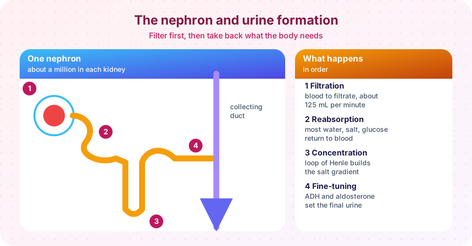
The working unit is the nephron. Each kidney has about a million, with the range in healthy adults roughly 0.5 to 1.5 million. A nephron is a long, thin tube, closed at one end, with a ball of capillaries pushed into the closed end.
Step 1: Filtration
Blood enters the glomerulus through a wide afferent arteriole and leaves by a narrower efferent arteriole. This arrangement keeps the pressure in the glomerular capillaries unusually high, about 55 mmHg. It pushes fluid out through a three-layered filtration barrier: the capillary wall with its pores, a basement membrane, and specialized cells called podocytes with slits between their "foot" processes. The barrier lets water, ions, glucose, amino acids and urea through but holds back blood cells and most proteins. The fluid collects in a cup around the glomerulus called Bowman's capsule.
The rate, the GFR, is about 125 mL per minute, or 180 liters per day. Doctors estimate GFR from blood creatinine, a waste product of muscle, because it is a good measure of overall kidney function. A falling GFR is the hallmark of chronic kidney disease. Finding protein or blood in urine is a sign that the filter is damaged, as normal glomeruli should hold both back.
The kidney protects its own GFR by autoregulation: if blood pressure rises or falls, the afferent arteriole narrows or widens so the flow through the glomerulus stays fairly steady across a wide range of arterial pressures (about 80 to 180 mmHg).
Step 2: Reabsorption in the proximal tubule
The filtrate flows into the proximal convoluted tubule (PCT), whose cells have a thick brush border like the intestine. This is where the bulk of reabsorption occurs. About 65 percent of the sodium and water, nearly all the glucose and amino acids, and much of the potassium and bicarbonate are returned to the blood. Sodium is pumped out of the tubule cells by the sodium-potassium pump, and water, glucose and amino acids follow in cotransport with sodium, in the same way as in the intestine.
Each transporter has a maximum capacity. If blood glucose is very high, as in uncontrolled diabetes, filtered glucose exceeds what the transporters can carry back, and some stays in the urine, carrying water with it. This is the renal threshold mentioned in the glucose lesson, and the reason why untreated diabetes causes thirst and frequent urination.
Step 3: The loop of Henle and the salt gradient
The tubule dips into the medulla as the loop of Henle and returns to the cortex. The two limbs behave differently. The descending limb is permeable to water but not to salt, so water leaves into the salty tissue. The ascending limb is impermeable to water but actively pumps salt out. This countercurrent arrangement, worked out by Werner Kuhn and others in the 1950s, builds up a gradient of salt concentration in the medulla, rising from about the same concentration as blood (300 mOsm/L) at the cortex to about four times that (up to 1,200 mOsm/L) deep in the medulla. Without this gradient the kidney could not make urine more concentrated than blood. Desert animals such as the kangaroo rat have extra-long loops, and can make extremely concentrated urine.
Step 4: Fine-tuning in the distal tubule and collecting duct
The last segments, the distal convoluted tubule (DCT) and the collecting duct, are where hormones decide the final result.
- ADH (antidiuretic hormone), released by the posterior pituitary when blood becomes too concentrated or blood volume falls, inserts water channels called aquaporins into the collecting-duct cells. Water then flows out into the salty medulla and into the blood, and a small volume of concentrated urine is produced. Without ADH, the collecting duct is nearly impermeable, and the person passes large volumes of dilute urine. Alcohol suppresses ADH release, which is why drinking alcohol makes you urinate more.
- Aldosterone, from the adrenal cortex, makes the distal tubule take back more sodium (and water follows) and secrete potassium.
- Atrial natriuretic peptide (ANP), released by the stretched atria of the heart, does the opposite: it increases sodium and water loss to lower blood volume.
Secretion also happens here, moving potassium, hydrogen ions, and many drugs from blood into the tubule. This is why the kidney can excrete acid and why doses of some medicines must be reduced when kidney function falls.
Controlling blood pressure: the renin-angiotensin system
Specialized cells next to the glomerulus, the juxtaglomerular cells, detect a fall in blood pressure or sodium delivery and release the enzyme renin. Renin converts a plasma protein into angiotensin I. An enzyme (ACE), found especially in lung capillaries, converts this to angiotensin II, a powerful vasoconstrictor that also stimulates aldosterone release and thirst. The result is higher blood pressure and a greater blood volume. Several widely used blood pressure medicines work by blocking steps in this chain; the details belong in a pharmacology course.
Storing and releasing urine
Urine flows through the ureters by peristalsis, not by gravity alone. The bladder can hold 400 to 600 mL, though the urge to urinate arises at roughly 200 to 300 mL. When it fills, stretch receptors send signals to the spinal cord and brain. The internal urethral sphincter (smooth muscle) opens automatically, while the external sphincter (skeletal muscle) is under voluntary control, and children learn to control it between about two and four years of age. The female urethra is only about 4 cm long and the male about 20 cm, which partly explains why bladder infections are more common in women.
Other kidney jobs, and what happens when they fail
Besides making urine, the kidneys produce erythropoietin and activate vitamin D, and they help maintain acid-base balance (see the next lesson). In kidney failure, wastes such as urea accumulate, fluid is retained, anemia develops and bones weaken. Dialysis can take over part of the work. The Dutch physician Willem Kolff built the first practical artificial kidney during the Second World War in occupied Netherlands, using sausage casing as a membrane. Hemodialysis passes blood past a membrane with fluid on the other side so wastes diffuse out. A kidney transplant is the best treatment for many patients.
Common misconceptions
"Urine is made by the bladder." The bladder only stores it. Urine is made in the nephrons.
"The kidneys get rid of toxins only when you drink more." Drinking enough water helps keep urine dilute, but the kidneys filter continuously, and extra water beyond need mainly increases urine volume.
"Clear urine means perfect health, dark urine means disease." Color mainly reflects dilution; foods, vitamins and medicines also change it. Blood or persistent changes need medical advice.
Try it
The first TryIt plots how glucose is filtered, reabsorbed and excreted as blood glucose rises. It is a simplified model, using a single transport maximum for all nephrons. In reality nephrons vary, so glucose appears in the urine at somewhat lower plasma levels (the "splay").
Press Run (or Ctrl + Enter) to see the result here.
Program input · what you'd type while the program runs
Type the answers before you press Run, one per line. Each time the program reads a line (with input()), it gets the next line from this box.
The second TryIt is a water-balance simulation: change how much water you have drunk and see how ADH and the urine respond. It is a qualitative model with invented constants.
Check your understanding
Quick check · 5 questions
of 5 answered You scored / 5 · perfect!
-
1. Where does most reabsorption of water, sodium and glucose occur?
About 65 percent of sodium and water and nearly all glucose and amino acids are reabsorbed in the proximal tubule. -
2. What does ADH do?
ADH inserts aquaporins into the collecting duct, letting water move out of the tubule into the blood. -
3. Why does a very high blood glucose cause glucose to appear in urine?
Reabsorption uses a limited number of carriers; when saturated, the excess is excreted. -
4. What is the function of the loop of Henle?
The countercurrent arrangement builds a salty medulla that draws water out of the collecting duct. -
5. Which sequence begins with a fall in blood pressure?
The renin-angiotensin-aldosterone system raises blood pressure and volume.
Summary
- The nephron filters blood at the glomerulus (about 125 mL/min), reabsorbs most of the filtrate and secretes extra solutes.
- The proximal tubule reabsorbs most solute and water; the loop of Henle builds a salt gradient; the distal tubule and collecting duct are controlled by aldosterone and ADH.
- The renin-angiotensin-aldosterone system raises blood pressure; ANP opposes it.
- The kidney also makes erythropoietin, activates vitamin D and, when it fails, can be replaced in part by dialysis or a transplant.
Track your progress
Create a free account to tick off lessons and pick up where you left off on any device.
of Anatomy and Physiology lessons completed