


















Lesson 33 of 52 · Respiratory and Digestive Systems
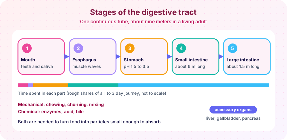
The digestive tract: from mouth to small intestine
Follow food from the mouth through the esophagus and stomach into the small intestine, and learn what acid, enzymes, bile and gut hormones do.
In 1822 a fur trader named Alexis St. Martin was accidentally shot in the stomach at a trading post in what is now Michigan. The wound healed but left an opening, or fistula, into his stomach. An army surgeon, William Beaumont, treated him and over the following years carried out experiments through the hole. He lowered pieces of food on a string into the stomach and watched what happened, and collected gastric juice. Beaumont proved that digestion in the stomach is chemical as well as mechanical and that the juice contains hydrochloric acid. The experiments would not meet modern ethical standards, since St. Martin had little choice, but they laid the foundation of gastric physiology.
This lesson follows a meal from the mouth to the end of the small intestine. It covers the structure of the gut wall, the mechanical and chemical steps in each region, the main enzymes, and the hormones that coordinate them. The next lesson finishes the story with absorption, the liver and the large intestine.
What you will learn
- Describe the four layers of the gut wall and the two kinds of gut movement.
- List the main steps of digestion in the mouth, esophagus, stomach and small intestine.
- Name the major digestive enzymes, what each digests and where it works.
- Explain what gastrin, secretin and cholecystokinin do.
- Explain why stomach acid does not digest the stomach itself.
Key terms
Alimentary canal: the continuous tube from mouth to anus.
Chyme: the semi-liquid mixture of food and gastric juice leaving the stomach.
Enzyme: a protein that speeds up a chemical reaction without being used up.
Bile: a fluid made by the liver that emulsifies fats; it contains no digestive enzymes.
Sphincter: a ring of muscle that opens and closes a passage.
The wall of the gut
From the esophagus to the anus the wall has the same four layers.
- The mucosa is the inner lining, with an epithelium that secretes mucus, enzymes or hormones, or absorbs.
- The submucosa is connective tissue with blood vessels, lymphatics and a nerve network.
- The muscularis externa has an inner ring of circular muscle and an outer layer of longitudinal muscle.
- The serosa is a smooth outer covering.
The muscle layers produce two kinds of movement. Peristalsis is a wave of contraction behind the food and relaxation ahead of it, pushing the contents forward. Segmentation, seen mainly in the small intestine, is a rhythmic pinching of the tube into segments that mixes the contents with enzymes and exposes them to the wall. The movements are directed by the enteric nervous system, a network of about 100 million neurons embedded in the gut wall that can run these reflexes without instructions from the brain. For that reason it is sometimes called the "second brain".
The mouth
Digestion begins before the first bite. In the early 1900s Ivan Pavlov, who received the 1904 Nobel Prize for work on digestive physiology, showed that the sight and smell of food make dogs salivate: a nervous reflex prepares the gut. In the mouth:
- Teeth grind food. Adults have up to 32 teeth: incisors cut, canines tear and molars crush.
- Saliva, from three pairs of salivary glands at roughly 1 to 1.5 liters a day, moistens food and contains salivary amylase, which splits starch into shorter sugars, and a little lingual lipase. It also contains mucus and antibacterial agents.
- The tongue shapes food into a bolus.
Swallowing starts voluntarily, then becomes a reflex. The soft palate closes the nasal passage, the larynx lifts and the epiglottis tips over the airway, and the bolus moves into the esophagus.
The esophagus
The esophagus is a muscular tube about 25 centimeters long. Peristalsis carries the bolus to the stomach in about 5 to 10 seconds; this is why you can swallow while hanging upside down. At the bottom the lower esophageal sphincter opens to let food in and then closes to stop acid from backing up. When it relaxes at the wrong time, acid reflux causes heartburn.
The stomach
The stomach is a J-shaped muscular bag that can hold about 1 to 1.5 liters comfortably. It stores food, starts protein digestion, kills many microbes and mixes the contents into chyme. Its wall has three layers of muscle (including an oblique one), so it can churn as well as squeeze.
The lining is dotted with gastric pits leading to glands with several cell types:
- Parietal cells secrete hydrochloric acid (HCl), making gastric juice very acidic, pH about 1.5 to 3.5, and intrinsic factor, which is needed to absorb vitamin B12 later in the small intestine.
- Chief cells secrete pepsinogen, an inactive precursor. Acid converts it to pepsin, an enzyme that cuts proteins into shorter chains. Releasing it in inactive form protects the cells that make it.
- Mucous cells secrete a thick alkaline mucus layer that shields the wall from acid and pepsin.
- G cells release the hormone gastrin into the blood, which stimulates more acid.
The acid unfolds proteins so that pepsin can reach them, kills many swallowed microbes and activates pepsin. A pH of 2 means about 100,000 times more hydrogen ions than a neutral pH 7, as you can calculate in the TryIt below.
In 1982 the Australian researchers Barry Marshall and Robin Warren found that most stomach ulcers are caused by the bacterium Helicobacter pylori, which colonizes the mucus layer, not by stress or spicy food as had been thought. Marshall famously drank a culture of the bacteria to demonstrate it. The pair shared the 2005 Nobel Prize in Physiology or Medicine, and ulcers are now usually treated with antibiotics plus acid reduction.
Food stays in the stomach for about two to four hours, depending on its fat content. Small squirts of chyme pass through the pyloric sphincter into the small intestine.
The small intestine
The small intestine is the main site of digestion and absorption. It is about 6 meters long in a living person, and has three parts: the duodenum (about 25 cm), the jejunum and the ileum. Most chemical digestion happens in the duodenum, where chyme meets secretions from the pancreas and liver.
The pancreas, liver and gallbladder
The pancreas releases pancreatic juice through a duct into the duodenum. It contains:
- bicarbonate, which neutralizes the acid in chyme so that pancreatic enzymes can work (they prefer a pH around 7 to 8),
- pancreatic amylase for starch,
- trypsin and chymotrypsin, proteases released as inactive precursors and activated in the gut,
- pancreatic lipase, the main fat-digesting enzyme,
- nucleases for DNA and RNA.
The liver makes bile, about 500 to 1,000 milliliters a day. Bile is stored and concentrated in the gallbladder and released into the duodenum when fat arrives. Bile salts do not digest anything chemically. They emulsify fat, breaking large droplets into tiny ones that give pancreatic lipase a much larger surface to act on, much like washing-up liquid breaks grease into droplets.
Enzymes on the brush border
The cells lining the small intestine have microscopic projections called microvilli, forming a "brush border." Enzymes anchored in it complete digestion: lactase, sucrase and maltase split disaccharides into monosaccharides; peptidases split small peptides into amino acids. Many adults lose much of their lactase activity after childhood, so lactose reaches the large intestine undigested, causing gas and discomfort. This is lactose intolerance, which is the usual state among adults in many parts of the world, while lactase persistence is common in populations with a long history of dairying.
| Nutrient | Mouth | Stomach | Small intestine | End product |
|---|---|---|---|---|
| Starch | Salivary amylase | (acid stops amylase) | Pancreatic amylase, brush-border enzymes | Glucose |
| Protein | None | Pepsin | Trypsin, chymotrypsin, peptidases | Amino acids |
| Fat | Little | Little | Bile and pancreatic lipase | Fatty acids and monoglycerides |
Hormonal control
Three hormones coordinate the process.
- Gastrin (stomach): released when food stretches the stomach; stimulates acid secretion.
- Secretin (duodenum): released when acid enters the duodenum; tells the pancreas to release bicarbonate. It was the first hormone discovered, by Bayliss and Starling in 1902, as you saw in the endocrine lesson.
- Cholecystokinin (CCK) (duodenum): released when fat and protein arrive; causes the gallbladder to contract and the pancreas to release enzymes, and signals fullness to the brain.
Common misconceptions
"Bile is a digestive enzyme." Bile contains salts and pigments, but no enzymes. It emulsifies fats; lipase does the digesting.
"Most digestion happens in the stomach." The stomach starts protein digestion, but most chemical digestion and nearly all absorption occur in the small intestine.
"Ulcers are caused by stress and spicy food." Most are caused by H. pylori or by long-term use of certain pain medicines.
Try it
First, the pH scale is logarithmic. This short program compares hydrogen ion concentrations along the digestive tract. The pH values are typical approximations.
Press Run (or Ctrl + Enter) to see the result here.
Program input · what you'd type while the program runs
Type the answers before you press Run, one per line. Each time the program reads a line (with input()), it gets the next line from this box.
Second, an enzyme sorter. Choose an enzyme and see the substrate and place of action. It is a study aid with simplified information.
Check your understanding
Quick check · 5 questions
of 5 answered You scored / 5 · perfect!
-
1. Which enzyme begins starch digestion in the mouth?
Salivary amylase starts splitting starch; pepsin and trypsin act on proteins. -
2. Why is pepsin released as pepsinogen?
Pepsinogen is inactive. Acid in the stomach lumen converts it, protecting the chief cells. -
3. What is the main function of bile?
Bile salts emulsify fat into small droplets, giving lipase more surface to work on. -
4. Which hormone tells the pancreas to release bicarbonate when acid enters the duodenum?
Secretin is released by the duodenum in response to acid and stimulates bicarbonate secretion. -
5. What does intrinsic factor do?
Parietal cells release intrinsic factor, which binds vitamin B12 so it can be absorbed in the ileum.
Summary
- The gut wall has mucosa, submucosa, muscle and serosa; peristalsis moves food and segmentation mixes it.
- Mechanical digestion (chewing, churning) and chemical digestion (acid, enzymes) work together.
- The stomach adds acid and pepsin; the pancreas adds bicarbonate, amylase, proteases and lipase; the liver adds bile.
- Brush-border enzymes complete digestion into monosaccharides, amino acids and fatty acids.
- Gastrin, secretin and CCK coordinate the secretions.
Track your progress
Create a free account to tick off lessons and pick up where you left off on any device.
of Anatomy and Physiology lessons completed