


















Lesson 13 of 52 · Skeletal and Muscular Systems
Bone structure, growth, remodeling and healing
Explore compact and spongy bone, osteons, osteoblasts and osteoclasts, how bones grow and remodel, how calcium is controlled and how fractures heal.
Pick up a skeleton in a classroom and it looks like chalk. In a living person, bone is nothing like chalk: it is warm, wet, richly supplied with blood vessels and nerves, and constantly being taken apart and rebuilt. Bone combines two opposite properties that engineers rarely get in one material. It is hard and resists compression, thanks to calcium salts, yet it is tough and resists cracking, thanks to flexible collagen. Pound for pound, that mix makes bone a remarkably light and durable structure.
This lesson opens bone up from the outside in: the gross structure of a long bone, the microscopic osteon, the four cell types, how bones form and grow, how remodeling controls blood calcium, and how a fracture heals.
What you will learn
- The parts of a long bone and the difference between compact and spongy bone.
- The microscopic structure of an osteon and the roles of the four bone cell types.
- How bones form (ossification) and lengthen at the growth plate.
- How remodeling and hormones keep blood calcium stable.
- The stages of fracture healing.
Key terms
- Diaphysis / epiphysis: the shaft / the expanded end of a long bone.
- Periosteum: the tough outer membrane that covers bone and carries blood vessels and nerves.
- Osteon: the basic structural unit of compact bone, a set of concentric rings around a central canal.
- Osteoblast / osteoclast: a cell that builds new bone / a cell that breaks bone down.
- Remodeling: the lifelong cycle of bone breakdown and rebuilding.
Classifying bones by shape
Bones come in five shapes that match their jobs. Long bones (femur, humerus, fingers) are longer than wide and act as levers. Short bones (carpals in the wrist, tarsals in the ankle) are cube-shaped and provide stability with limited movement. Flat bones (skull, sternum, ribs, scapula) are thin and protect organs or offer broad surfaces for muscles. Irregular bones (vertebrae, facial bones) have complex shapes. Sesamoid bones form inside tendons; the kneecap (patella) is the largest.
Anatomy of a long bone
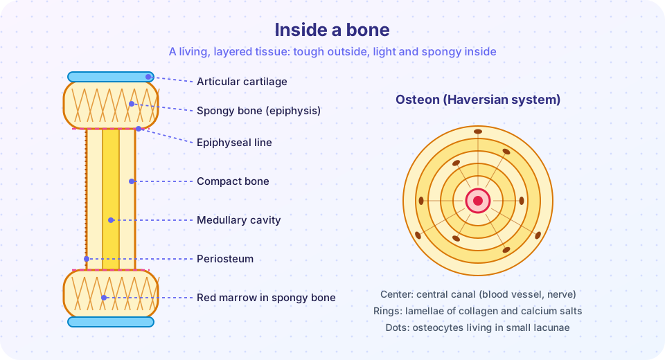
A typical long bone, such as the femur, has these parts:
- The diaphysis is the long shaft, a tube of dense bone.
- The two epiphyses are the expanded ends, covered by smooth articular cartilage where they meet other bones, which cushions and reduces friction.
- The metaphysis is the flared region between each epiphysis and the diaphysis; in a growing child it contains the epiphyseal plate (growth plate) of cartilage, and in an adult, the epiphyseal line, the bony remnant.
- The periosteum covers the outside (except the cartilage-covered ends). Its outer layer is tough fibrous tissue, and its inner layer holds bone-forming cells. Tendons and ligaments blend into it, and its blood vessels feed the bone.
- The medullary cavity in the shaft is a hollow filled with yellow marrow (fat) in adults, and the thin endosteum lines it.
- The epiphyses contain spongy bone with red marrow, which makes blood cells.
Bone has two textures. Compact bone is dense and smooth and forms the outer shell, making up about 80% of the skeleton's mass. Spongy (trabecular) bone is a lattice of thin plates called trabeculae with spaces filled with marrow. The trabeculae are not random: they line up along the directions of stress, which is a clever design that gives strength with minimum weight.
Microscopic structure: the osteon
Under a microscope, compact bone is made of cylinders called osteons (Haversian systems), each about 0.2 millimeters wide, running mostly along the bone's length. Each osteon has:
- A central (Haversian) canal carrying blood vessels and nerves.
- Concentric rings called lamellae, layers of matrix in which collagen fibers run in alternating directions, like plywood.
- Small cavities called lacunae between lamellae, each housing an osteocyte (a mature bone cell).
- Tiny channels called canaliculi that link lacunae to each other and to the central canal, so osteocytes can exchange nutrients and wastes through a network of cell extensions.
- Perforating (Volkmann's) canals, which run crosswise and connect the blood supply of neighboring osteons and the periosteum.
The matrix of bone is about 65% inorganic mineral (mostly hydroxyapatite, a calcium phosphate crystal) and about 35% organic material (mainly collagen). Calcium salts supply hardness and the collagen supplies tensile strength and flexibility. Remove the mineral with acid and a bone becomes rubbery and can be tied in a knot; remove the collagen by heating and it becomes brittle and crumbles.
The four bone cells
| Cell | Job |
|---|---|
| Osteoprogenitor cell | stem cell in the periosteum and endosteum that becomes an osteoblast |
| Osteoblast | builds new bone matrix; secretes collagen and promotes mineral deposition |
| Osteocyte | mature cell inside a lacuna; maintains the matrix and senses mechanical stress |
| Osteoclast | large multinucleate cell that dissolves bone with acid and enzymes (resorption) |
How bones form and grow
The skeleton starts as soft tissue and hardens in a process called ossification. In intramembranous ossification, flat bones of the skull and most of the clavicle form directly inside sheets of embryonic connective tissue. In endochondral ossification, which builds most other bones, a small model of hyaline cartilage is first laid down and gradually replaced by bone, starting in the middle of the shaft (the primary ossification center) around the eighth week of fetal life. Secondary ossification centers appear in the epiphyses, mostly after birth.
Lengthening happens at the epiphyseal plate. Cartilage cells on the epiphysis side divide and push the ends apart, while cartilage on the shaft side is replaced by bone. Growth in width happens by adding bone beneath the periosteum. Growth hormone, thyroid hormone and, in puberty, sex hormones drive this. Eventually, in the late teens to early twenties, the plate is completely replaced by bone (the plate "closes") and the bone can no longer grow longer. Doctors can estimate the age of a child by checking which growth plates are open on an X-ray.
Remodeling and calcium
Bone is never finished. Osteoclasts and osteoblasts work as teams in a cycle: osteoclasts dissolve a small patch, then osteoblasts fill it with new bone. Each year about 5% to 10% of the adult skeleton is remodeled, so the entire skeleton is renewed roughly every ten years or so, although the rate differs between bone types. Remodeling has two purposes:
- Mechanical adaptation. Bone strengthens where it is loaded and weakens where it is not, an observation called Wolff's law (after Julius Wolff, 1892). Weight-bearing exercise builds bone, while bed rest, space flight and paralysis cause bone loss.
- Calcium homeostasis. Bones hold about 99% of the body's calcium. Blood calcium is held in a narrow range, about 2.2 to 2.6 millimoles per liter (roughly 9 to 10.5 mg per 100 mL), because calcium is essential for nerve signals, muscle contraction and blood clotting. When blood calcium falls, the parathyroid glands release parathyroid hormone (PTH), which stimulates osteoclasts to release calcium from bone, tells the kidneys to retain calcium and increases activation of vitamin D, which raises calcium absorption from the gut. When calcium is high, PTH falls and more calcium is deposited in bone. This is a negative feedback loop, the key homeostatic design you will meet again in Chapter 8.
The simulation below is a toy version of that loop. It is a teaching model with made-up units: it shows only that a hormone responding to a shortfall brings the level back toward the set point, not real hormone values.
Press Run (or Ctrl + Enter) to see the result here.
Program input · what you'd type while the program runs
Type the answers before you press Run, one per line. Each time the program reads a line (with input()), it gets the next line from this box.
Another hormone, calcitonin from the thyroid, can lower blood calcium by inhibiting osteoclasts, but in adult humans its role is small compared with PTH. Vitamin D is also needed so that the gut can absorb calcium, which is why a child who gets too little vitamin D can develop soft, bowed bones, a condition called rickets. In adults the equivalent is osteomalacia.
Fractures and healing
A fracture is a break in a bone. Doctors classify them as closed (simple) if the skin is intact and open (compound) if a bone end pierces the skin, which carries a risk of infection. Other descriptions include transverse, spiral, comminuted (broken into several pieces) and greenstick (an incomplete break in a child's flexible bone). Healing follows predictable stages:
- Hematoma (hours to days). Torn vessels bleed and a blood clot forms around the break.
- Fibrocartilaginous (soft) callus (about 1 to 3 weeks). Fibroblasts and chondroblasts invade the clot and produce collagen and cartilage, which bridges the gap.
- Bony (hard) callus (about 3 to 4 months). Osteoblasts replace the cartilage with new spongy bone.
- Remodeling (months to years). Osteoclasts and osteoblasts reshape the area, and compact bone replaces the excess spongy bone until the bone resembles its original form.
A cast or pin holds the broken ends still so that the callus can form. These timings are approximate and depend on the bone, the person's age, nutrition and blood supply.
Bone health across life
Bone mass builds during childhood and adolescence and peaks around the late twenties to early thirties. After that it slowly declines. Osteoporosis ("porous bone") is a condition of low bone density that makes fractures more likely. It is more common in older women, partly because the fall in estrogen after menopause speeds up resorption. Habits that favor strong bones include weight-bearing and resistance exercise, enough calcium and vitamin D in the diet, no smoking and moderate alcohol. This is general educational information; individual advice belongs with a health professional.
Common misconceptions
- "Bones stop changing after you stop growing." They remodel for life.
- "Drinking lots of milk guarantees strong bones." Calcium helps, but vitamin D, exercise, hormones and genetics matter as well.
- "A broken bone heals back with a scar like skin." Bone heals with new bone, and a good repair can leave almost no trace.
- "Yellow marrow makes blood." Red marrow does. Yellow marrow is mostly fat, though it can convert to red marrow when needed.
Check your understanding
Quick check · 5 questions
of 5 answered You scored / 5 · perfect!
-
1. Which cell type dissolves bone matrix?
Osteoclasts are large cells that resorb bone and release calcium. -
2. What do canaliculi do?
Canaliculi are tiny channels linking osteocytes in lacunae to each other and to the central canal. -
3. Blood calcium drops. What happens next?
PTH rises when calcium is low, and it stimulates osteoclasts, kidney retention and vitamin D activation. -
4. At which site does a child's long bone increase in length?
Cartilage cells in the epiphyseal plate divide and are replaced by bone, lengthening the shaft. -
5. Which sequence of fracture healing is correct?
A clot forms first, then a cartilage callus, then a bony callus, then remodeling.
Summary
- Long bones have a shaft, two ends, a periosteum and a marrow cavity; compact bone forms the shell and spongy bone fills the ends.
- Compact bone is built of osteons with central canals, lamellae, lacunae and canaliculi; the matrix is about 65% mineral and 35% collagen.
- Osteoblasts build, osteocytes maintain and osteoclasts resorb bone; remodeling lasts a lifetime.
- Bones form by intramembranous or endochondral ossification and lengthen at the epiphyseal plate until it closes.
- PTH, vitamin D and mechanical load regulate bone; fractures heal through hematoma, soft callus, hard callus and remodeling.
Track your progress
Create a free account to tick off lessons and pick up where you left off on any device.
of Anatomy and Physiology lessons completed