


















Lesson 14 of 52 · Skeletal and Muscular Systems
The skeleton and joints: bones, articulations and movement
Learn the 206 bones of the axial and appendicular skeleton, the spine and ribs, the three joint classes, synovial joint types and movement terms.
Count the bones in an adult and you get 206, though the exact number depends on who counts, because some people have extra small bones in the hands, feet or spine and some bones are fused. A newborn has about 270 to 300 bones, many of them partly cartilage; as children grow, several bones fuse together, for example the five sacral bones that fuse into one sacrum.
The sheer number of bones is less important than the pattern. The skeleton has two main divisions, and the places where bones meet, the joints or articulations, decide how much the skeleton can move. This lesson gives you the map of the skeleton, the three classes of joints and the vocabulary of movement.
What you will learn
- The two divisions of the skeleton and the main bones in each.
- The regions and curves of the vertebral column and the types of ribs.
- The three structural classes of joints and the parts of a synovial joint.
- The six types of synovial joint and the standard terms for movements.
Key terms
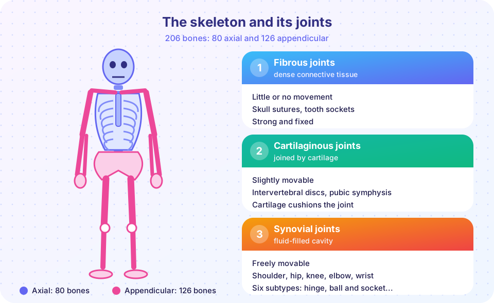
- Axial skeleton: the skull, vertebral column and thoracic cage (80 bones).
- Appendicular skeleton: the limbs and the girdles that attach them to the axial skeleton (126 bones).
- Joint (articulation): a place where two or more bones meet.
- Synovial joint: a freely movable joint with a fluid-filled cavity.
- Range of motion: how far a joint can move in each direction.
The axial skeleton
The skull has 22 bones, in two groups. Eight cranial bones form the protective case around the brain: the frontal, two parietal, two temporal, occipital, sphenoid and ethmoid bones. Fourteen facial bones form the face: two maxillae (upper jaw), two zygomatic (cheekbones), two nasal, two lacrimal, two palatine, two inferior nasal conchae, the vomer and the mandible (lower jaw). The mandible is the only skull bone that moves freely; the others are locked together by immovable joints called sutures. Inside the middle ears are three tiny bones per ear (the malleus, incus and stapes, 6 in total) that carry sound, and the hyoid, a U-shaped bone in the neck that anchors the tongue, floats without joining any other bone.
The vertebral column (spine) has 26 bones in an adult: 7 cervical vertebrae in the neck, 12 thoracic vertebrae that carry ribs, 5 lumbar vertebrae in the lower back, 1 sacrum (five fused vertebrae) and 1 coccyx (the tailbone, three to five fused). Between adjacent vertebrae lie intervertebral discs of fibrocartilage with a gel-like center that absorb shock; a "slipped" disc means the center has bulged out and may press on a nerve. Seen from the side, the spine has four gentle curves (cervical, thoracic, lumbar and sacral) that act as a spring, spreading out forces from walking and lifting. The first two cervical vertebrae are special: the atlas (C1) holds up the skull and allows nodding "yes", while the axis (C2) has a peg called the dens around which the atlas rotates, allowing a shake of the head "no".
The thoracic cage consists of 12 pairs of ribs, 12 thoracic vertebrae and the sternum (breastbone). The sternum has three parts: the manubrium, the body and the xiphoid process. The upper seven pairs of ribs, the true ribs, attach directly to the sternum through costal cartilage; pairs 8 to 10 (false ribs) attach indirectly; pairs 11 and 12 (floating ribs) do not attach at the front. The cage protects the heart and lungs and flexes slightly to let you breathe.
Counting: skull 22 + ear bones 6 + hyoid 1 + vertebral column 26 + sternum 1 + ribs 24 = 80 axial bones.
The appendicular skeleton
The pectoral (shoulder) girdle attaches each arm to the trunk. It has two bones on each side: the clavicle (collarbone) at the front and the scapula (shoulder blade) at the back. The only bony attachment to the axial skeleton is where each clavicle meets the sternum, which gives the shoulder great freedom of movement but also makes it easy to dislocate.
Each upper limb has 30 bones: the humerus in the upper arm; the radius (on the thumb side) and ulna (on the little-finger side) in the forearm; 8 carpals in the wrist; 5 metacarpals in the palm; and 14 phalanges in the fingers (two in the thumb and three in each of the other four fingers).
The pelvic girdle is made of two hip bones, each formed by the fusion of the ilium, ischium and pubis. With the sacrum and coccyx, it makes the pelvis, a strong basin that carries the weight of the upper body and protects the pelvic organs. The female pelvis is typically wider and shallower with a larger opening, which suits childbirth.
Each lower limb has 30 bones: the femur (thigh bone, the longest and strongest bone in the body), the patella (kneecap), the tibia (shin bone, which carries the weight) and the thin fibula (on the outer side of the leg); 7 tarsals in the ankle and heel; 5 metatarsals in the foot; and 14 phalanges in the toes.
Counting: shoulder girdles 4 + upper limbs 60 + hip bones 2 + lower limbs 60 = 126 appendicular bones, so 80 + 126 = 206. The program below does that arithmetic and draws the split.
Press Run (or Ctrl + Enter) to see the result here.
Program input · what you'd type while the program runs
Type the answers before you press Run, one per line. Each time the program reads a line (with input()), it gets the next line from this box.
Joints: classification by structure
A joint is any place where bones meet. They can be classed by structure (what holds the bones together) or by function (how much movement they allow). The two schemes match closely:
| Structural class | Held together by | Function | Examples |
|---|---|---|---|
| Fibrous | dense connective tissue | synarthrosis: immovable | skull sutures, tooth in socket, tibia-fibula ends |
| Cartilaginous | cartilage | amphiarthrosis: slightly movable | intervertebral discs, pubic symphysis, growth plates |
| Synovial | fluid-filled joint cavity | diarthrosis: freely movable | shoulder, hip, knee, elbow, wrist |
Newborns have gaps between skull bones called fontanelles ("soft spots") that allow the head to be compressed during birth and the brain to grow; they close over about the first 18 to 24 months.
Anatomy of a synovial joint
Synovial joints have a standard set of parts:
- Articular cartilage (hyaline) covers the ends of the bones, reducing friction.
- The joint (articular) capsule is a sleeve of fibrous connective tissue around the joint, lined by a synovial membrane.
- The synovial fluid secreted by that membrane lubricates, nourishes the cartilage and absorbs shock. It is thick, like egg white, which is where the name comes from (syn "together", ovum "egg").
- Ligaments reinforce the capsule and limit movement in unwanted directions.
- Some have menisci (pads of fibrocartilage, as in the knee) and bursae, small fluid-filled sacs that reduce friction where tendons or skin pass over bone. Inflammation of a bursa is bursitis.
The knee is a good case study. Four main ligaments keep the femur and tibia in line: the anterior cruciate ligament (ACL) prevents the tibia from sliding forward, the posterior cruciate (PCL) prevents it from sliding back, and the two collateral ligaments resist sideways forces. The two menisci cushion and deepen the joint. A sudden twist can tear the ACL, a common sports injury.
Six types of synovial joint
| Type | Movement | Example |
|---|---|---|
| Plane (gliding) | sliding in one plane | between carpals, between vertebral processes |
| Hinge | bend and straighten in one plane | elbow, knee, finger joints |
| Pivot | rotation around a long axis | atlas and axis, radius and ulna |
| Condyloid (ellipsoid) | two planes, no rotation | wrist (radius and carpals), knuckles |
| Saddle | two planes with a wider range | thumb's base (carpometacarpal) |
| Ball-and-socket | all planes, including rotation | shoulder, hip |
The language of movement
Movements are described relative to anatomical position.
- Flexion decreases the angle between bones (bending the elbow); extension increases it (straightening). Going beyond the normal straight position is hyperextension. At the ankle, lifting the foot upward is dorsiflexion and pointing the toes down is plantar flexion.
- Abduction moves a limb away from the midline (raising the arm sideways); adduction brings it toward the midline.
- Rotation turns a bone around its own long axis; circumduction moves a limb in a cone, as in drawing a circle with your arm.
- Pronation rotates the forearm so the palm faces backward (or down); supination turns the palm forward (or up). A mnemonic: supination is the "soup" position, holding a bowl of soup in the palm.
- Inversion turns the sole of the foot inward; eversion turns it outward.
- Elevation and depression raise and lower a body part, such as shrugging the shoulders or opening the jaw.
The widget below shows a simple hinge joint. It is a diagram model of the elbow, with the range limited to about 0 to 145 degrees as in a typical adult, rather than a medical measurement tool.
Common joint problems
A sprain is a stretched or torn ligament, and a strain is an injured muscle or tendon. A dislocation is when the bones of a joint are forced out of alignment. Arthritis means joint inflammation. Osteoarthritis is the common "wear and tear" type in which articular cartilage thins with age and use. Rheumatoid arthritis is an autoimmune disease in which the immune system attacks the synovial membrane. Treatments vary and belong in clinical care; here the point is that structure explains the symptoms.
Common misconceptions
- "Joints are where bones touch." In synovial joints the bones are separated by cartilage and a film of fluid, not in direct contact.
- "Cracking your knuckles causes arthritis." Studies have not found that habit to cause arthritis; the sound comes from gas bubbles in synovial fluid.
- "Humans have 206 bones at every age." Babies have more; fusion reduces the number.
- "The clavicle is a minor bone." It is the only bony strut linking the arm to the trunk, so it breaks easily in falls.
Check your understanding
Quick check · 6 questions
of 6 answered You scored / 6 · perfect!
-
1. Which group of bones belongs to the appendicular skeleton?
The scapula and clavicle form the pectoral girdle, part of the appendicular skeleton. -
2. How many cervical vertebrae are there?
There are 7 cervical, 12 thoracic and 5 lumbar vertebrae, plus the sacrum and coccyx. -
3. Which joint type allows rotation in all planes?
Ball-and-socket joints, such as the shoulder and hip, move in all planes. -
4. Skull sutures are classified as which type of joint?
Sutures are fibrous joints joined by dense connective tissue and are immovable. -
5. Moving your arm away from the midline sideways is called what?
Abduction takes a limb away from the midline; adduction brings it toward the midline. -
6. What is the job of synovial fluid?
Synovial fluid reduces friction, absorbs shock and feeds the cartilage.
Summary
- The adult skeleton has 206 bones: 80 axial (skull, spine, thoracic cage) and 126 appendicular (girdles and limbs).
- The spine has 26 bones in five regions and four curves; ribs include true, false and floating pairs.
- Joints are fibrous (immovable), cartilaginous (slightly movable) or synovial (freely movable).
- Synovial joints have cartilage, a capsule, fluid and ligaments; they come in six types.
- Movement terms such as flexion, extension, abduction and rotation are defined from anatomical position.
Next: Muscle contraction
Track your progress
Create a free account to tick off lessons and pick up where you left off on any device.
of Anatomy and Physiology lessons completed