


















Lesson 20 of 52 · Nervous System and Senses
The brain and spinal cord: regions, protection and pathways
Tour the CNS: cerebral lobes, thalamus, hypothalamus, brainstem and cerebellum, plus meninges, cerebrospinal fluid and the spinal cord with its tracts.
The human brain weighs about 1.3 to 1.4 kilograms, has the consistency of soft tofu and contains roughly 86 billion neurons. It is protected by a thick skull, three membranes and a cushion of fluid, because it cannot repair itself the way skin can, and because it is the one organ you cannot live without for more than a few minutes. Yet it is also strikingly organized: different regions specialize in different tasks, and the way they connect is as important as what each one does.
This lesson tours the central nervous system (CNS) from the outside in. You will see the protective layers, then the brain's major regions and their functions, with the landmark cases that taught us what they do, and finally the spinal cord, the brain's two-way highway to the body.
What you will learn
- How the skull, meninges, cerebrospinal fluid and blood-brain barrier protect the CNS.
- The main parts of the brain and what each does, with key evidence.
- The structure of the spinal cord, its tracts and its connections to the body.
- The idea of localization of function and its limits.
Key terms
- Cerebral cortex: the thin outer layer of gray matter of the cerebrum, where most conscious processing happens.
- Gray matter / white matter: areas rich in neuron cell bodies / areas of myelinated axons.
- Meninges: three protective membranes (dura mater, arachnoid mater, pia mater) around the brain and spinal cord.
- Cerebrospinal fluid (CSF): clear fluid that cushions the CNS and fills its internal spaces.
- Tract: a bundle of axons in the CNS with a shared origin and destination.
Protecting the CNS
The CNS has four layers of defense.
- Bone. The skull encloses the brain and the vertebral column encloses the spinal cord.
- Meninges. Three membranes lie between bone and nervous tissue: the tough dura mater (outermost), the web-like arachnoid mater and the delicate pia mater, which clings to the brain's surface. Bleeding between these layers, for example after a head blow, can compress the brain, and infection of them is called meningitis.
- Cerebrospinal fluid. CSF fills the subarachnoid space (between arachnoid and pia) and four internal chambers called ventricles. It is made by capillary networks called the choroid plexuses at a rate of about 500 milliliters per day, while the total volume at any moment is only about 150 milliliters, so it is replaced several times a day. CSF cushions the brain (the brain "floats", which reduces its effective weight to a few dozen grams), removes waste and helps keep the chemical environment steady.
- The blood-brain barrier (BBB). Capillaries in the brain have unusually tight junctions between their cells, supported by astrocyte extensions. Oxygen, carbon dioxide, glucose and lipid-soluble substances such as alcohol pass, while many bacteria, toxins and most large drug molecules do not. The barrier makes brain drug treatment a challenge.
The cerebrum
The cerebrum is the largest part of the brain, divided into left and right cerebral hemispheres by a deep groove and linked by a thick band of about 200 million axons, the corpus callosum. The wrinkled surface is the cerebral cortex, 2 to 4 millimeters of gray matter. Folding packs a large area into a small skull: the folds are gyri (ridges) and the grooves sulci. Beneath the cortex is white matter, and deeper still are clusters of gray matter called the basal nuclei, which help to start and smooth movements. In Parkinson's disease and Huntington's disease these circuits are affected.
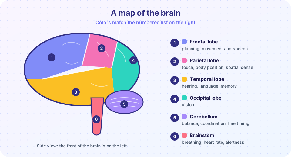
Each hemisphere has four lobes, named after the skull bones above them (see the picture above):
- Frontal lobe (1): planning, decision-making, personality, voluntary movement and, usually in the left hemisphere, speech production. At its back lies the primary motor cortex (the precentral gyrus), which sends commands to skeletal muscles. The motor map of the body is distorted: areas with fine control, such as the hands and mouth, get a large share of cortex, a picture known as the "homunculus" from the work of the neurosurgeon Wilder Penfield in the 1930s to 1950s.
- Parietal lobe (2): touch, body position and spatial awareness. The primary somatosensory cortex (the postcentral gyrus), just behind the motor strip, receives touch, pressure, temperature and pain signals.
- Temporal lobe (3): hearing, language comprehension and memory. The primary auditory cortex lies here, and the hippocampus, tucked inside, is vital for forming new memories.
- Occipital lobe (4): vision. Damage to the primary visual cortex causes blindness even when the eyes are healthy.
Language illustrates localization with nuance. In 1861 the French physician Paul Broca described a patient nicknamed "Tan" who understood speech but could say almost nothing. After his death a lesion was found in the left frontal lobe, now called Broca's area, a region important for producing speech. In 1874 Carl Wernicke described a different pattern in patients with fluent but meaningless speech and poor comprehension, tied to the left temporal lobe (Wernicke's area). In most people, language depends mainly on the left hemisphere, in about 95% of right-handed and about 70% of left-handed people. Still, modern imaging shows language uses a wide network. The popular idea that people are "left-brained" (logical) or "right-brained" (creative) is a myth; the hemispheres cooperate in almost every task.
The famous cases
Two cases from the history of neurology show how damage reveals function, though case reports are limited, because one person cannot show how every brain works. Phineas Gage, a railroad worker, survived an iron rod through his frontal lobe in 1848 and was reported to have changed personality, which drew attention to the frontal lobe's role in behavior. The details of how much he changed have been debated, and later accounts exaggerated them. Henry Molaison ("H.M."), who had parts of both temporal lobes, including the hippocampus, removed in 1953 to stop severe seizures, lost the ability to form new long-term memories of facts and events while his skills (such as a mirror-drawing task, studied by Brenda Milner) could still improve, showing that different memory types depend on different structures.
The diencephalon, brainstem and cerebellum
Beneath the cerebrum lies the diencephalon. The thalamus (egg-shaped, paired) is the sensory relay station: nearly all sensory signals except smell pass through it on their way to the cortex. The hypothalamus, only the size of an almond, is the master regulator of homeostasis: it controls body temperature, hunger, thirst, sleep cycles and emotions, and it commands the pituitary gland, linking the nervous and endocrine systems (Chapter 5).
The brainstem connects the brain to the spinal cord and has three parts. The midbrain controls eye movement and visual and auditory reflexes. The pons (Latin for bridge) relays signals between the cerebrum and cerebellum and helps regulate breathing. The medulla oblongata houses the vital centers that control heartbeat, blood pressure, breathing and reflexes such as coughing, swallowing and vomiting. Within the brainstem, a diffuse network called the reticular formation controls wakefulness and arousal. Damage to the brainstem is often fatal because of the vital centers there.
The cerebellum ("little brain") sits at the back and underneath, and contains about 80% of the brain's neurons in a region only about 10% of its volume. It does not start movements; it refines them by comparing the intended movement with the actual position of the body and correcting errors, giving you balance, posture and smooth, precisely timed motion. Cerebellar damage produces clumsy, uncoordinated movement.
The limbic system, a set of structures that includes the hippocampus (memory) and the amygdala (emotion, especially fear), connects the cortex with the hypothalamus and links thoughts, emotions and body responses.
Try the explorer below. It is a study aid that links each region to its main functions and to a classic example of what happens when the region is damaged.
The spinal cord
The spinal cord is a cylinder of nervous tissue about the thickness of a finger and 42 to 45 centimeters long. It runs from the base of the brainstem through the vertebral canal and ends around the level of the first or second lumbar vertebra (L1 to L2) in the conus medullaris. Below that, spinal nerve roots hang down like a horse's tail, the cauda equina, which is why a doctor can safely take a CSF sample with a needle below that level (a lumbar puncture, at about L3 to L4).
In cross section, the cord shows a butterfly-shaped core of gray matter surrounded by white matter. The posterior (dorsal) horns of gray matter receive sensory information, the anterior (ventral) horns contain the cell bodies of motor neurons that supply skeletal muscle, and small lateral horns in the thoracic and upper lumbar regions contain autonomic neurons. The white matter consists of tracts: ascending tracts carry sensory information up to the brain (for example the dorsal columns for fine touch and body position, and the spinothalamic tracts for pain and temperature), and descending tracts carry motor commands down (the corticospinal tract controls voluntary movement). Many tracts cross over (decussate), so the left side of the brain largely controls the right side of the body.
Thirty-one pairs of spinal nerves leave the cord. Each attaches by two roots: the dorsal root brings in sensory fibers, whose cell bodies lie in the dorsal root ganglion, and the ventral root carries motor fibers out. The two roots merge into one mixed nerve. The cord also works on its own: it coordinates reflexes without involving the brain, which you will study in the next lesson. Because the cord is a communication cable, injuries at different levels cause loss of movement and sensation below the level of injury.
The program below counts spinal nerve pairs by region and draws the pattern. The 31 pairs are 8 cervical, 12 thoracic, 5 lumbar, 5 sacral and 1 coccygeal. It also does the CSF turnover arithmetic.
Press Run (or Ctrl + Enter) to see the result here.
Program input · what you'd type while the program runs
Type the answers before you press Run, one per line. Each time the program reads a line (with input()), it gets the next line from this box.
Common misconceptions
- "The brain is a muscle." It is nervous tissue. Using it does not make it grow bigger like a muscle, although learning changes synapses.
- "People are left-brained or right-brained." Research shows the two hemispheres work together; language is lateralized, but personality types are not.
- "The brain is fixed after childhood." It remains plastic throughout life, though it changes more slowly.
- "CSF is just water." It is a controlled fluid with a distinct composition, made from blood plasma and low in protein.
Check your understanding
Quick check · 6 questions
of 6 answered You scored / 6 · perfect!
-
1. Which lobe contains the primary visual cortex?
The occipital lobe at the back of the brain processes vision. -
2. Which structure relays sensory information to the cortex?
The thalamus is the relay station for most sensory input. -
3. What is the order of the meninges from the skull inward?
Dura mater is outermost, then arachnoid mater, then pia mater on the brain surface. -
4. Where do the cell bodies of sensory neurons that enter the spinal cord lie?
Sensory neuron cell bodies are in the dorsal root ganglion, just outside the cord. -
5. What did the case of H.M. reveal?
After removal of his hippocampi, H.M. could not form new declarative memories. -
6. Which part of the brain houses the vital centers for breathing and heart rate?
The medulla has cardiovascular and respiratory centers essential for life.
Summary
- The CNS is protected by bone, three meninges, cerebrospinal fluid and the blood-brain barrier.
- The cerebrum has four lobes (frontal, parietal, temporal, occipital) with specialized areas; the corpus callosum links the hemispheres.
- The thalamus relays sensory input, the hypothalamus regulates homeostasis, the brainstem controls vital functions and the cerebellum coordinates movement.
- The spinal cord has a gray matter core and white matter tracts; 31 pairs of spinal nerves attach through dorsal (sensory) and ventral (motor) roots.
- Function is localized, but behavior always arises from networks of regions.
Track your progress
Create a free account to tick off lessons and pick up where you left off on any device.
of Anatomy and Physiology lessons completed