


















Lesson 8 of 52 · Organization of the Body
Cell structure, organelles and the cell cycle
Tour a human cell: membrane, nucleus, mitochondria, ER, Golgi and lysosomes, see how DNA becomes protein, and follow the cell cycle and mitosis.
In 1665 Robert Hooke looked at a slice of cork through an early microscope and saw rows of tiny boxes, which he named cells, after the cells of a monastery. By the 1830s Matthias Schleiden and Theodor Schwann proposed that all plants and animals are made of cells, and in 1855 Rudolf Virchow added that every cell comes from another cell. Together these ideas form the cell theory: cells are the basic unit of life, all organisms are made of cells, and cells arise only from existing cells.
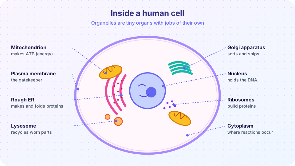
A human cell is a busy factory the size of a speck of dust. It takes in raw materials, builds products, recycles waste and, when the time comes, copies itself. This lesson tours the factory floor: the outer membrane, the control office (nucleus), the power plants (mitochondria), the assembly lines and the delivery system.
What you will learn
- The structure of the plasma membrane and the main organelles with their functions.
- How the cytoskeleton gives cells shape and movement.
- How a gene becomes a protein (transcription and translation).
- The stages of the cell cycle and of mitosis.
Key terms
- Organelle: a specialized structure inside a cell with a particular job.
- Cytosol / cytoplasm: the jelly-like fluid inside the cell / that fluid plus all the organelles.
- Gene: a stretch of DNA that carries the instructions for a protein or functional RNA.
- Mitosis: division of the nucleus that gives two genetically identical daughter nuclei.
- Chromosome: a tightly packed molecule of DNA with its proteins; humans have 46 in most body cells.
The plasma membrane
The plasma membrane is the flexible boundary that separates the inside of the cell from its surroundings. In 1972 S. Jonathan Singer and Garth Nicolson described it with the fluid mosaic model: a double layer of phospholipids (the bilayer) in which proteins float like boats on a sea. The phospholipid heads face the watery inside and outside; the oily tails face each other in the middle. This arrangement forms by itself, because it hides the tails from water.
Embedded in the bilayer are proteins that act as channels, carriers, receptors that catch hormones and chemical signals, enzymes and markers that identify the cell as "self". Cholesterol tucked between phospholipids keeps the membrane flexible but not too runny. Short sugar chains on the outer surface form the glycocalyx, which helps cells recognize each other. The membrane is selectively permeable: it lets some things pass freely and blocks others. The next lesson shows how.
The cytoplasm and cytoskeleton
Inside the membrane lies the cytoplasm: the cytosol (mostly water with dissolved ions, sugars and proteins) plus the organelles. The cytoskeleton is a network of protein filaments that works as scaffolding, rails and muscle all at once. There are three kinds:
- Microfilaments (actin, the thinnest) support the cell's shape and drive movement such as muscle contraction and cell crawling.
- Intermediate filaments are rope-like and resist stretching. Keratin filaments make skin cells tough.
- Microtubules (the thickest) are hollow tubes made of tubulin. They hold organelles in place, form tracks for motor proteins that haul cargo, build the spindle that pulls chromosomes apart, and form the core of cilia and flagella.
Hair-like cilia line the airways and sweep mucus; a single flagellum drives a sperm cell; microvilli are tiny finger-like folds that increase surface area in the intestine and kidney.
The nucleus and ribosomes
The nucleus is the control center. A double membrane, the nuclear envelope, surrounds it, studded with pores that let messenger molecules through. Inside, DNA is wrapped around proteins and loosely arranged as chromatin; when a cell prepares to divide, the chromatin condenses into visible chromosomes. The nucleolus, a dense spot, builds the parts of ribosomes. Most human cells have one nucleus; mature red blood cells have none, and skeletal muscle fibers have many.
Ribosomes are tiny molecular machines made of RNA and protein that build proteins by reading messenger RNA. Some float free in the cytosol (making proteins used inside the cell), and others sit on the endoplasmic reticulum (making proteins to be exported or placed in membranes).
The protein assembly line
Proteins are made and shipped through connected compartments, called the endomembrane system:
- The rough endoplasmic reticulum (rough ER) is a network of flattened sacs with ribosomes on its surface. It folds new proteins and packs them into small bubbles called vesicles.
- The smooth ER has no ribosomes. It makes lipids and steroids, stores calcium (in muscle) and detoxifies drugs, which is why liver cells are rich in it.
- The Golgi apparatus, a stack of flattened sacs like pancakes, modifies, sorts and packages proteins into vesicles labeled for delivery to the membrane or outside the cell.
- Lysosomes are vesicles filled with digestive enzymes that break down worn-out organelles, bacteria and debris. If lysosomal enzymes are missing, waste builds up, as in Tay-Sachs disease. Peroxisomes break down fatty acids and neutralize hydrogen peroxide.
Mitochondria: the power plants
Mitochondria make most of the cell's ATP through aerobic respiration, using oxygen to extract energy from glucose and fats. Each has a smooth outer membrane and an inner membrane folded into cristae, which increase the surface area where ATP is made. Cells with high energy needs, such as heart muscle, have thousands of mitochondria. Mitochondria also contain their own small circular DNA, which is inherited almost entirely from the mother, and divide on their own. Together with their bacteria-like size, this supports the endosymbiotic theory (proposed by Lynn Margulis in 1967): mitochondria descend from bacteria that took up residence inside ancestral cells about two billion years ago.
From gene to protein
A cell's DNA holds about 20,000 protein-coding genes. Making a protein takes two steps. In transcription, inside the nucleus, an enzyme copies a gene into a molecule of messenger RNA (mRNA). The mRNA leaves through a nuclear pore. In translation, a ribosome reads the mRNA three letters (one codon) at a time, and for each codon a transfer RNA delivers the matching amino acid, building the chain. The start codon is AUG (which codes for methionine), and three codons (UAA, UAG, UGA) mean stop. The code is nearly universal across life.
The program below is a simplified teaching model of these two steps. It transcribes a short DNA strand into mRNA and then translates it with the standard genetic code. Real cells add many extra steps, but the logic matches.
Press Run (or Ctrl + Enter) to see the result here.
Program input · what you'd type while the program runs
Type the answers before you press Run, one per line. Each time the program reads a line (with input()), it gets the next line from this box.
The cell cycle and mitosis
Cells grow and divide through a repeating sequence called the cell cycle. Most of the time is spent in interphase, when the cell does its normal work and gets ready to divide:
- G1 (first gap): the cell grows and makes proteins and organelles.
- S (synthesis): DNA is copied, so each chromosome now consists of two identical sister chromatids joined at a centromere.
- G2 (second gap): the cell checks the copies and finishes preparing.
Then comes mitosis, the division of the nucleus, in four stages, followed by cytokinesis, the division of the cytoplasm:
- Prophase: chromosomes condense and become visible; the nuclear envelope breaks down; the spindle of microtubules forms.
- Metaphase: chromosomes line up along the cell's middle.
- Anaphase: the sister chromatids separate and are pulled to opposite poles.
- Telophase: nuclear envelopes re-form around the two sets and the chromosomes loosen; the cell pinches in two.
The result is two genetically identical daughter cells, each with 46 chromosomes. A typical cultured human cell takes about 24 hours for the whole cycle, with mitosis lasting only about an hour. Cell division varies a lot across the body: cells lining the intestine divide every few days, whereas most mature neurons and heart muscle cells rarely or never divide. Sperm and egg cells are made by a different process, meiosis, which halves the chromosome number to 23 (Chapter 7).
Cells must also be able to die in an orderly way. Apoptosis is programmed cell death, a tidy self-dismantling that sculpts fingers from webbed paddles in the embryo and removes damaged cells. Cancer is, in simple terms, a failure of the controls on division and death: cells divide when they should not and ignore the signals to stop.
Why cells are small
Cells are microscopic for a good physical reason: they need to exchange materials across the membrane, which grows with surface area, while their needs grow with volume. Doubling a cell's radius multiplies its surface by 4 but its volume by 8. The plot below shows the surface-area-to-volume ratio of a sphere, 3 / r, as the radius grows.
Press Run (or Ctrl + Enter) to see the result here.
Program input · what you'd type while the program runs
Type the answers before you press Run, one per line. Each time the program reads a line (with input()), it gets the next line from this box.
Common misconceptions
- "Mitochondria create energy." Energy cannot be created. Mitochondria transfer energy from food molecules to ATP.
- "The nucleus is the whole cell's brain and the cell cannot live without it." Control comes from DNA, but red blood cells live about 120 days with no nucleus at all.
- "Mitosis is the whole cell cycle." It is the short division phase; most of the cycle is interphase.
- "Every cell has 46 chromosomes." Most body cells do, but egg and sperm cells have 23, and red blood cells have none.
Check your understanding
Quick check · 6 questions
of 6 answered You scored / 6 · perfect!
-
1. Which part of the cell controls what enters and leaves?
The plasma membrane is selectively permeable and controls traffic into and out of the cell. -
2. Which sequence correctly shows the path of a protein made for export?
Proteins for export are made on the rough ER, modified in the Golgi and carried to the membrane in vesicles. -
3. In which phase of the cell cycle is DNA replicated?
S stands for synthesis: the DNA is copied during this phase of interphase. -
4. What happens during anaphase?
Anaphase is when the sister chromatids are pulled apart. -
5. Why does a doubling of cell radius cause problems for exchange?
Surface area scales with the square of the radius, volume with the cube, so the surface-to-volume ratio falls. -
6. Which molecule carries the copy of a gene from the nucleus to a ribosome?
Messenger RNA is made in transcription and read by the ribosome in translation.
Summary
- The fluid mosaic membrane controls entry and exit; the cytoskeleton gives shape, rails and movement.
- The nucleus holds DNA; ribosomes, rough ER and Golgi build and ship proteins; lysosomes recycle; mitochondria make ATP.
- Genes are transcribed to mRNA and translated into proteins at the ribosome.
- The cell cycle has G1, S, G2 and mitosis (prophase, metaphase, anaphase, telophase) plus cytokinesis.
- Small cells exchange materials efficiently because of their high surface-to-volume ratio.
Next: Membrane transport
Track your progress
Create a free account to tick off lessons and pick up where you left off on any device.
of Anatomy and Physiology lessons completed