


















Lesson 31 of 52 · Respiratory and Digestive Systems
Breathing and lung mechanics
Learn the airways, how the diaphragm and ribs create pressure changes that move air, what surfactant does, and how lung volumes are measured.
You breathe about 20,000 times a day, and most of the time you never think about it. Breathing, or pulmonary ventilation, is not done by the lungs themselves. The lungs contain no muscle. They are elastic bags that are stretched open and allowed to spring back by the muscles and bones around them. The principle that makes this work was set out by the Irish chemist Robert Boyle in 1662: for a fixed amount of gas at constant temperature, pressure and volume are inversely related. Change the volume of the chest and you change the pressure of the air inside it, and air flows from higher pressure to lower pressure.
This lesson follows the airways from nose to alveoli, explains how the diaphragm and ribs make air move, describes the role of surfactant and the pleural membranes, and introduces the lung volumes that a spirometer measures. It also describes how the brain sets the pace of breathing.
What you will learn
- Trace air through the conducting airways to the alveoli.
- Explain inspiration and expiration using Boyle's law, the diaphragm and the intercostal muscles.
- Describe the roles of the pleura and surfactant.
- Define tidal volume, vital capacity and the other standard lung volumes.
- Explain how the medulla and chemoreceptors regulate breathing.
Key terms
Pulmonary ventilation: the movement of air into and out of the lungs.
Diaphragm: the dome-shaped skeletal muscle below the lungs that is the main muscle of inspiration.
Pleura: the double membrane around each lung, with a thin film of fluid between the layers.
Surfactant: a mixture of lipids and proteins that lowers surface tension in the alveoli.
Tidal volume: the volume of air moved in a single quiet breath.
The airways
Air enters through the nose or mouth. The nasal cavity warms, moistens and filters it: coarse hairs catch large particles and a mucus lining traps smaller ones. The air then passes the pharynx (throat) and the larynx, or voice box, which holds the vocal cords and is guarded by the epiglottis. Below the larynx is the trachea, a tube about 10 to 12 centimeters long held open by 16 to 20 C-shaped rings of cartilage. The open part of each C faces the esophagus, so a swallowed lump of food can bulge into the trachea's soft back wall without catching.
The trachea splits into the right and left primary bronchi, which divide again and again, about 23 times in total, into ever finer bronchioles. This branching, whose pattern resembles an upside-down tree, ends in clusters of microscopic air sacs, the alveoli. An adult has roughly 300 to 500 million of them. The airways down to the terminal bronchioles are the conducting zone: they only carry air and do no gas exchange. The volume of air in them, about 150 mL, is called anatomical dead space.
The airway lining is a layer of cells with cilia, microscopic hairs, and mucus-producing goblet cells. Cilia beat in coordinated waves and carry mucus and trapped dust upward toward the throat, where it is swallowed or coughed out. This "mucociliary escalator" is damaged by smoking, which is one reason why smokers cough.
Why the lungs follow the chest
Each lung is wrapped in a thin double-layered membrane, the pleura. One layer sticks to the lung surface and the other lines the inside of the chest wall. Between them is a thin film of fluid, pleural fluid, which acts like water between two glass plates: the layers slide easily but cannot be pulled apart. The lung therefore follows the chest wall when it expands. The pressure in the pleural space (intrapleural pressure) is slightly lower than atmospheric pressure, about -4 mmHg relative to the air at rest, which keeps the lung inflated. If air enters the pleural space through a wound or a leak in the lung (a pneumothorax), the seal is lost and the lung partly collapses.
Inspiration
Quiet inspiration is an active process that uses muscles:
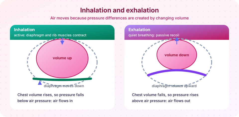
- The diaphragm, a dome of skeletal muscle, contracts and flattens, moving downward. It is driven by the phrenic nerve, which arises from the third to fifth cervical nerves in the neck. A spinal injury above this level can stop breathing, which is why a mnemonic is taught to students: "C3, 4, 5 keeps the diaphragm alive".
- The external intercostal muscles between the ribs contract and lift the ribs up and out, like the handle of a bucket swinging up.
- The volume of the chest cavity rises in every direction.
- By Boyle's law the pressure inside the lungs, the alveolar pressure, falls a little below atmospheric pressure, to about -1 mmHg relative to the outside. Air flows in until the pressures are equal.
The pressure difference is tiny. Only 1 or 2 mmHg is enough to move half a liter of air in about two seconds.
Expiration
Quiet expiration is mostly passive. The muscles relax, and the elastic tissue of the lungs and chest wall recoils. Volume falls, alveolar pressure rises to about +1 mmHg, and air flows out. When you breathe out forcefully, for example when blowing out candles or exercising hard, the internal intercostals and the abdominal muscles contract and squeeze the air out faster.
Surfactant and compliance
The inner surface of each alveolus is coated with a thin film of water. Water molecules attract each other strongly, which creates surface tension that tends to collapse the alveolus. Specialized cells called type II alveolar cells secrete surfactant, which interrupts the attraction between water molecules and cuts surface tension. Without it, breathing would take enormous effort. Babies born very early may not yet make enough, causing respiratory distress syndrome; treatment with artificial surfactant, introduced in the 1980s, has greatly improved survival.
Compliance is how easily the lungs stretch. Conditions that stiffen the lungs, such as fibrosis, reduce compliance and make breathing harder. Elastic recoil, in contrast, is how well the lungs spring back. Emphysema destroys elastic fibers, so air is trapped and expiration becomes difficult.
Lung volumes
A spirometer records the volume of air moved in and out. The values below are approximate averages for a healthy young adult. They vary with size, age, sex and fitness.
| Volume | Approx. size | Meaning |
|---|---|---|
| Tidal volume (TV) | 500 mL | A normal quiet breath |
| Inspiratory reserve volume (IRV) | 3,000 mL | Extra air you can breathe in after a quiet inspiration |
| Expiratory reserve volume (ERV) | 1,100 mL | Extra air you can breathe out after a quiet expiration |
| Residual volume (RV) | 1,200 mL | Air that stays in the lungs even after maximal expiration |
| Vital capacity (VC) | about 4,600 mL | IRV + TV + ERV: the most you can breathe out after a full breath in |
| Total lung capacity (TLC) | about 5,800 mL | VC + RV |
Residual volume cannot be exhaled, so you cannot measure it directly with simple spirometry. Minute ventilation is tidal volume times breathing rate: 500 mL × 12 breaths per minute is 6 L/min. Because 150 mL of each breath stays in the dead space, alveolar ventilation is lower: (500 - 150) × 12 = 4.2 L/min. This is why slow, deep breaths are more effective for gas exchange than quick, shallow ones: the same minute ventilation delivers more fresh air to the alveoli.
Controlling breathing
The rhythm is generated in the medulla oblongata in the brainstem, where groups of neurons fire in a pattern that triggers the diaphragm roughly every four to five seconds. The pons smooths the transitions. Cerebral control lets you hold your breath, speak or sing, but the automatic centers take over when you sleep.
The most powerful drive to breathe is not lack of oxygen but rising carbon dioxide. Carbon dioxide in blood forms acid, and central chemoreceptors in the medulla detect the resulting change in pH of the surrounding fluid. Peripheral chemoreceptors in the carotid and aortic bodies respond to low oxygen, high carbon dioxide and low pH, and send signals to the medulla. This is why you feel the urge to breathe when you hold your breath: carbon dioxide is building up, not oxygen running out. Breathing a lot beforehand (hyperventilation) lowers carbon dioxide and delays the urge, which is dangerous in water because oxygen can fall to a level that causes unconsciousness before you feel the need to breathe.
Common misconceptions
"The lungs push air out." The lungs have no muscle. The diaphragm and rib muscles change chest volume, and the lungs follow.
"We breathe because oxygen runs low." In a healthy person the main trigger is a rise in carbon dioxide (a fall in pH).
"We use all the oxygen we breathe in." Inhaled air is about 21 percent oxygen and exhaled air about 16 percent; you extract only about a quarter of it at rest, which is why mouth-to-mouth rescue breathing works.
Try it
The first demonstration draws a spirogram, the trace a spirometer makes, from the typical volumes above. The curve is a schematic, not a recording of a real person.
Press Run (or Ctrl + Enter) to see the result here.
Program input · what you'd type while the program runs
Type the answers before you press Run, one per line. Each time the program reads a line (with input()), it gets the next line from this box.
The second demonstration is a Boyle's law calculator and a ventilation comparison. It uses fixed textbook numbers, so treat it as a model.
Check your understanding
Quick check · 5 questions
of 5 answered You scored / 5 · perfect!
-
1. What happens to the pressure inside the lungs when the diaphragm contracts and the chest volume increases?
By Boyle's law, more volume means lower pressure, so air flows in until pressures equalize. -
2. What is the role of surfactant?
Surfactant from type II alveolar cells interrupts water's cohesion, so the alveoli do not collapse. -
3. Which volume cannot be breathed out even after a maximal expiration?
Residual volume stays in the lungs after maximal expiration, keeping the alveoli open. -
4. A person breathes 15 times per minute with a tidal volume of 400 mL and 150 mL of dead space. What is the alveolar ventilation?
(400 - 150) mL × 15 = 3,750 mL/min = 3.75 L/min. -
5. What is the main normal trigger of the urge to breathe?
Chemoreceptors respond chiefly to carbon dioxide and pH; a fall in oxygen matters mainly when it becomes severe.
Summary
- Air moves because muscles change the chest volume and Boyle's law turns the volume change into a pressure change.
- Inspiration is active (diaphragm and external intercostals); quiet expiration is passive elastic recoil.
- The pleura couples the lungs to the chest wall; surfactant prevents alveolar collapse.
- Tidal volume, reserve volumes, residual volume and vital capacity describe the lungs' capacity; alveolar ventilation excludes dead space.
- The medulla sets the rhythm and chemoreceptors adjust it, mainly according to carbon dioxide.
Track your progress
Create a free account to tick off lessons and pick up where you left off on any device.
of Anatomy and Physiology lessons completed