


















Lesson 39 of 52 · Urinary and Reproductive Systems
Reproductive anatomy and gametes
Learn the organs of the male and female reproductive systems, how meiosis makes gametes, and what hormones control sperm and egg production.
Every other organ system exists to keep one body alive. The reproductive system exists to pass on genes. It is silent in childhood, begins to work at puberty and, in females, ends its reproductive function at menopause. Its central job is to make gametes, the egg and the sperm, which each carry half the genetic information of a person. When they fuse at fertilization, the full number is restored and a new individual begins.
This lesson describes the organs of the typical male and female reproductive systems, explains how meiosis shuffles genes, and compares the very different ways in which sperm and eggs are produced. It is an educational overview of typical anatomy and physiology; human bodies and development vary, and this lesson is not medical advice.
What you will learn
- Name the main organs of the female and male reproductive systems and their functions.
- Explain how meiosis differs from mitosis and why it increases variation.
- Compare spermatogenesis and oogenesis.
- Describe hormonal control of the testes and ovaries.
- Explain how sex is determined genetically and how development can vary.
Key terms
Meiosis: cell division that halves the chromosome number to make gametes.
Spermatogenesis: the production of sperm in the testes.
Oogenesis: the production of eggs (ova) in the ovaries.
Follicle: an ovarian structure containing a developing egg.
Gonadotropins: the pituitary hormones FSH and LH that stimulate the gonads.
Making gametes: meiosis
Most body cells have 46 chromosomes, arranged in 23 pairs: one chromosome of each pair from each parent. They divide by mitosis, producing two identical copies. Gametes are made by meiosis, a special division with two rounds that produces cells with only 23 chromosomes, one from each pair.
Meiosis creates variation in two ways. First, during the first division matching chromosomes exchange segments (crossing over), producing new combinations of genes. Second, the pairs are sorted into gametes independently (independent assortment): there are 2²³, about 8.4 million, possible combinations of whole chromosomes in a single person's gametes, before crossing over is counted. This is why siblings, other than identical twins, are genetically different. At fertilization, egg and sperm fuse to restore 46 chromosomes.
Sex chromosomes work like this. The typical pattern is two X chromosomes (XX) in females and an X and a Y (XY) in males. A gene on the Y chromosome called SRY, identified in 1990, triggers the embryonic gonad to develop into a testis; without it the gonad usually develops into an ovary. Development of the sex organs depends on many genes and hormones, and variations in sex development occur in a small proportion of people. Textbook descriptions describe the most common pattern.
The male reproductive system
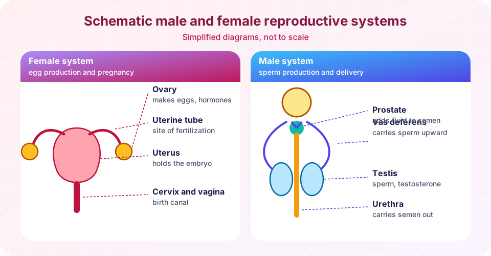
- Testes. Two oval organs, each about 4.5 cm long, hang in the scrotum, which keeps them about 2 to 3 °C below core body temperature, the temperature at which sperm production works best. Inside each testis are tightly coiled seminiferous tubules, where sperm are made, and Leydig cells between the tubules, which make testosterone.
- Epididymis. A long coiled tube on the back of each testis where sperm mature and are stored.
- Vas deferens. A muscular tube that carries sperm from the epididymis up into the pelvis. A vasectomy is the surgical cutting of this tube.
- Accessory glands. The seminal vesicles, prostate and bulbourethral glands add fluid that nourishes and carries the sperm. Sperm plus these fluids make semen. The seminal vesicles contribute fructose as fuel; the prostate adds an alkaline fluid.
- Urethra and penis. The urethra runs through the penis. It carries semen out, and also urine at other times; a sphincter prevents both from passing together.
Spermatogenesis
Sperm production begins at puberty and normally continues for life. Stem cells in the wall of the seminiferous tubules divide, mature through meiosis and take about 64 to 74 days to become sperm, with another week or two in the epididymis. A sperm cell is built for one job: a head holding the nucleus, topped by an acrosome of enzymes, a middle piece packed with mitochondria for energy, and a long tail (flagellum) for swimming. A healthy adult makes millions of new sperm each day.
Hormonal control. The hypothalamus releases GnRH in pulses, which makes the anterior pituitary release LH and FSH. LH stimulates the Leydig cells to make testosterone. FSH acts on Sertoli cells, the supporting cells in the tubules that nourish developing sperm. Testosterone and a hormone called inhibin, made by Sertoli cells, then feed back negatively on the hypothalamus and pituitary, a loop that keeps levels stable. Testosterone is also responsible for the male reproductive tract, for body hair, a deeper voice and muscle mass during puberty.
The female reproductive system
- Ovaries. Two almond-sized organs, each about 3 cm long, that release eggs and make estrogen and progesterone.
- Uterine (fallopian) tubes. Each tube, about 10 cm long, reaches from near an ovary to the uterus. It has finger-like fimbriae that sweep the released egg in, and cilia and muscle waves carry it along. Fertilization usually occurs here.
- Uterus. A hollow, thick-walled, pear-shaped organ of about 7 cm in length in the pelvis. Its wall has an outer connective layer, a thick middle layer of smooth muscle (the myometrium) and an inner lining, the endometrium, which thickens each cycle and is shed if no pregnancy occurs.
- Cervix and vagina. The cervix is the narrow lower end of the uterus; the vagina is a muscular canal that receives sperm and is the birth canal.
- Mammary glands. The breasts are modified sweat glands that produce milk after childbirth under the control of prolactin and oxytocin.
Oogenesis
Egg production differs markedly from sperm production. A female fetus forms all her primary oocytes before birth, about 1 to 2 million. They start meiosis and then pause. By puberty only about 300,000 to 400,000 remain, and over a lifetime about 400 to 500 are released. Each month a few follicles begin to grow under the influence of FSH, usually one becomes dominant and releases its egg at ovulation. Meiosis is completed only after fertilization.
In sperm production, one stem cell gives four equal sperm. In egg production, each meiotic division is uneven: the egg receives nearly all the cytoplasm and the other products, the polar bodies, are tiny and degenerate. The egg is the largest human cell, about 0.1 mm across, just visible to the naked eye, and carries the nutrients the early embryo needs.
The supply of follicles declines steadily with age. When it becomes low, typically around age 51 on average, the ovaries make much less estrogen and cycles stop. This is menopause, and it is defined as 12 consecutive months without a period. Unlike in the male, in whom sperm production continues with age (though it may decline), the female reproductive years are limited by this finite supply.
Hormones at puberty
Puberty starts when the hypothalamus increases GnRH pulses, which are low during childhood. LH and FSH then rise and stimulate the gonads to make sex steroids. Their effects include growth of the gonads and genital organs, and the secondary sex characteristics: breast development, widening of the hips and fat distribution in females; facial hair, voice deepening and greater muscle mass in males. The timing varies widely and has been shifting earlier in many countries, a trend linked to better nutrition and other factors.
Common misconceptions
"Women lose a single egg each month." A cohort of follicles starts to develop each cycle; usually one releases an egg, and the others degenerate.
"Sperm are made continuously but eggs are made monthly." Eggs begin development before birth; what happens monthly is the completion of growth and release of one.
"The Y chromosome alone makes someone male." A gene (SRY) on the Y starts testis development, but many other genes and hormones take part in sexual development.
Try it
The first TryIt shows the decline in the number of eggs through life, using approximate estimates from the literature. The curve is a schematic, plotted on a log scale, and individual numbers vary a great deal.
Press Run (or Ctrl + Enter) to see the result here.
Program input · what you'd type while the program runs
Type the answers before you press Run, one per line. Each time the program reads a line (with input()), it gets the next line from this box.
The second TryIt is a gamete shuffler. It shows three chromosome pairs, one colored by parent, and produces random gametes by independent assortment. It is a simplified demonstration of the idea; real meiosis involves 23 pairs and crossing over.
Check your understanding
Quick check · 5 questions
of 5 answered You scored / 5 · perfect!
-
1. How many chromosomes does a human gamete contain?
Meiosis halves the chromosome number from 46 to 23; fertilization restores 46. -
2. Which cells of the testis make testosterone?
Leydig cells, stimulated by LH, make testosterone. Sertoli cells nourish developing sperm and respond to FSH. -
3. Where does fertilization usually take place?
The egg and sperm usually meet in the upper part of the uterine tube. -
4. Which statement about oogenesis is correct?
A female fetus forms her primary oocytes, which start meiosis and pause; the supply declines with age. -
5. Why are the testes in the scrotum?
Sperm production works best at about 2 to 3 °C below core temperature.
Summary
- Gametes are made by meiosis, which halves the chromosome number and shuffles genes by crossing over and independent assortment.
- The male system makes sperm in the seminiferous tubules and delivers them through the epididymis, vas deferens and urethra, with fluid from accessory glands.
- The female system releases eggs from the ovaries into the uterine tubes, where fertilization occurs, and supports pregnancy in the uterus.
- GnRH, LH and FSH control the gonads; testosterone and estrogen feed back on the hypothalamus and pituitary.
- Sperm production is continuous after puberty; the supply of eggs is finite and falls with age.
Track your progress
Create a free account to tick off lessons and pick up where you left off on any device.
of Anatomy and Physiology lessons completed