


















Lesson 15 of 52 · Skeletal and Muscular Systems
Muscle contraction: the sliding filament model
Follow a contraction from the motor neuron signal to calcium release, the cross-bridge cycle and sarcomere shortening, then twitches, tetanus and motor units.
When you decide to lift a cup, a signal travels from your brain down a motor neuron and arrives at a muscle fiber, and within about a hundredth of a second thousands of tiny molecular motors start pulling. You feel the result as a smooth movement, yet it is built from events at a scale of nanometers. The understanding of how it works is one of the most elegant stories in physiology, and it is also a good example of structure explaining function.
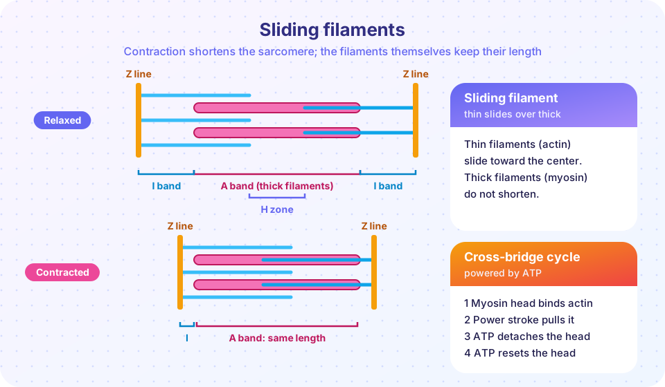
Two research teams solved the central puzzle in 1954. Andrew Huxley and Rolf Niedergerke in Cambridge, and Hugh Huxley and Jean Hanson at MIT, independently showed that muscle shortens when thin filaments slide past thick filaments, without either kind of filament changing its length. That idea, the sliding filament model, is still the basis of every textbook. This lesson builds it up from the whole muscle down to the molecules.
What you will learn
- The structure of a skeletal muscle from the whole muscle to the sarcomere.
- How a signal from a motor neuron crosses the neuromuscular junction.
- The steps of excitation-contraction coupling and the cross-bridge cycle.
- How twitches add up to smooth force, and what a motor unit is.
Key terms
- Sarcomere: the contractile unit of a muscle fiber, from one Z line to the next.
- Actin / myosin: the thin filament protein / the thick filament motor protein.
- Neuromuscular junction: the synapse between a motor neuron and a muscle fiber.
- Motor unit: one motor neuron and all the muscle fibers it controls.
- Cross-bridge: the temporary link formed when a myosin head attaches to actin.
From muscle to sarcomere
A whole skeletal muscle is wrapped in layers of connective tissue. The epimysium surrounds the entire muscle. Inside, the perimysium wraps bundles of fibers called fascicles, and each individual muscle fiber is wrapped by a delicate endomysium. These layers come together at the ends of the muscle to form the tendon, which carries the pull to the bone. A muscle fiber is a single, long, multinucleate cell, 10 to 100 micrometers across and as long as the muscle in some cases. Its plasma membrane is called the sarcolemma and its cytoplasm the sarcoplasm.
Each fiber is packed with rod-like myofibrils, about 1 micrometer across, made of repeating units, the sarcomeres, lined up end to end. Under a microscope the regular arrangement produces the dark and light striations that give skeletal muscle its name "striated". In each sarcomere:
- Z lines mark the ends.
- Thin filaments, made mainly of the protein actin, are anchored to the Z lines. Two other proteins sit on them: tropomyosin, a thread that lies along actin and covers its binding sites, and troponin, a small complex that holds tropomyosin in place and binds calcium.
- Thick filaments, made of bundles of the protein myosin, are in the middle. Each myosin molecule has a long tail and a head that sticks out like an oar and can bind to actin.
- The dark A band is the full length of the thick filaments (it does not change). The light I band is the part with thin filaments only. The H zone is the central region of the A band with thick filaments only. The M line is the center.
A relaxed sarcomere is about 2.5 micrometers long, so a single centimeter of muscle contains about 4,000 sarcomeres in series. When every sarcomere shortens by a little, the whole muscle shortens by a lot.
The nerve signal: the neuromuscular junction
Skeletal muscle never contracts by itself. It waits for a motor neuron, a nerve cell whose cell body lies in the spinal cord or brainstem and whose axon runs out to the muscle. The axon branches and ends on each fiber at a specialized synapse, the neuromuscular junction (NMJ):
- A nerve action potential reaches the axon terminal.
- Voltage-gated calcium channels open, and Ca²⁺ flows into the terminal.
- Vesicles of the neurotransmitter acetylcholine (ACh) fuse with the membrane (exocytosis) and release ACh into the narrow gap, the synaptic cleft.
- ACh binds to receptors on the muscle fiber's motor end plate. These receptors are channels that let sodium in, which depolarizes the sarcolemma and triggers a muscle action potential.
- An enzyme, acetylcholinesterase, breaks down ACh within a millisecond or two, ending the signal, so that one nerve impulse makes one twitch.
The NMJ is a target of several well-known substances. Botulinum toxin blocks ACh release (used in tiny, medically supervised doses to treat muscle spasms), curare blocks the receptor, and in the autoimmune disease myasthenia gravis the body makes antibodies against ACh receptors, causing muscle weakness. These are examples of how a step in the pathway explains a symptom, not medical guidance.
Excitation-contraction coupling
The action potential spreads along the sarcolemma and dives into the fiber through T tubules, narrow tunnels of membrane that reach deep into the cell. Next to each T tubule lies the sarcoplasmic reticulum (SR), a specialized smooth ER that stores calcium ions at a high concentration. The T tubule's voltage signal opens calcium release channels in the SR, and Ca²⁺ floods into the sarcoplasm around the myofibrils. This link between the electrical signal and mechanical contraction is called excitation-contraction coupling.
At rest, tropomyosin blocks the myosin-binding sites on actin. When Ca²⁺ binds to troponin, the troponin complex changes shape and tugs tropomyosin aside, exposing the binding sites. Now myosin heads can grab on.
The cross-bridge cycle
Each myosin head works like a tiny rower, repeating four steps:
- Attach. A myosin head, already "cocked" and carrying ADP and phosphate from earlier ATP splitting, binds to a site on actin, forming a cross-bridge.
- Power stroke. The head releases the phosphate and ADP and pivots, pulling the thin filament toward the center of the sarcomere by about 10 nanometers.
- Detach. A new ATP molecule binds to the myosin head, which releases it from actin.
- Reset ("cock"). ATP is split into ADP and phosphate, and the energy returns the head to its cocked, high-energy position, ready to bind again further along.
Each cycle takes a few milliseconds, and a muscle contraction involves billions of heads repeating the cycle asynchronously, so the filament moves smoothly, like many people pulling on a rope hand over hand. ATP is needed at two points: for detachment and for resetting. That explains rigor mortis: after death, ATP runs out, so myosin heads cannot detach from actin and the muscles stiffen for a day or so, until the proteins break down.
Relaxation happens when the nerve stops firing. ACh is cleared, the sarcolemma stops being excited, calcium pumps (which use ATP) return Ca²⁺ to the SR, troponin lets go and tropomyosin covers the actin binding sites again. The thin filaments slide back passively.
The widget below calculates the sarcomere's band lengths as it shortens, using typical filament lengths (thick filaments about 1.6 µm, thin filaments about 1.0 µm each side). It shows that the A band never changes while the I band and H zone shrink, and it looks up the relative force the sarcomere can produce at that length.
Motor units, twitches and graded force
A single motor neuron branches to supply anywhere from a few to a thousand or more fibers, and the neuron plus its fibers is a motor unit. When the neuron fires, all its fibers contract together. Muscles that need fine control, such as those moving the fingers or eyes, have small motor units (a handful of fibers per neuron), while the large muscles of the back and legs have big ones.
A single action potential in a fiber produces a brief twitch: a short delay (latent period), a rise in force and a decline as calcium is pumped back. A twitch lasts about 10 to 100 milliseconds, faster in the eye muscles and slower in the calf. If a second stimulus arrives before the muscle has fully relaxed, the second twitch builds on the first. This is wave summation. At high stimulation rates the individual twitches blend into a smooth, sustained contraction, fused tetanus, which is the normal way your muscles work. (This tetanus is only the physiological word for a sustained contraction; it has no connection to the infection tetanus.)
The force of a muscle is graded in two ways: by changing the rate of firing (summation) and by changing the number of active motor units, a process called recruitment. Small, easily excited motor units are recruited first and big ones later, so force increases smoothly (Henneman's size principle). An individual fiber, however, follows the all-or-none rule: when it is stimulated above threshold it contracts fully.
The simulation below adds idealized twitches. It is a mathematical toy, with made-up time constants and a saturation rule, but it shows the three regimes: separate twitches at low rates, summation at moderate rates and a fused plateau at high rates.
Press Run (or Ctrl + Enter) to see the result here.
Program input · what you'd type while the program runs
Type the answers before you press Run, one per line. Each time the program reads a line (with input()), it gets the next line from this box.
Length and tension
How hard a muscle can pull depends on its starting length, because that sets how many cross-bridges can form. At about 2.0 to 2.25 µm sarcomere length, the thin filaments overlap almost all of the myosin heads and tension is greatest. If the muscle is overstretched, the filaments barely overlap and there are few heads to pull; if it is too short, the thin filaments collide and crowd, and the force falls. In the body, resting muscle length sits near the best range, which is why the shape of the length-tension curve matters for exercise technique and for the best positions for strength.
Press Run (or Ctrl + Enter) to see the result here.
Program input · what you'd type while the program runs
Type the answers before you press Run, one per line. Each time the program reads a line (with input()), it gets the next line from this box.
Types of contraction
In an isometric contraction the muscle produces force without changing length, as when you push against a wall. In an isotonic contraction it changes length while the load stays about the same. A concentric contraction shortens the muscle (lifting a cup), and an eccentric contraction lengthens it while it still produces force (lowering the cup slowly). Eccentric work is gentler on the energy budget but more likely to cause the delayed muscle soreness that follows unfamiliar exercise.
Common misconceptions
- "Muscle filaments shorten." Neither actin nor myosin changes length. The sarcomere shortens because the filaments slide.
- "Muscles need ATP only to contract." They need ATP to detach and reset the myosin heads and to pump calcium back. That is why a muscle with no ATP is stiff in rigor, not relaxed.
- "Calcium comes from the bloodstream during contraction." It is released from the sarcoplasmic reticulum inside the fiber.
- "A bigger twitch means a bigger signal." A single fiber is all-or-none; force is graded by firing rate and by recruiting more fibers.
Check your understanding
Quick check · 6 questions
of 6 answered You scored / 6 · perfect!
-
1. Which band stays the same length when a sarcomere shortens?
The A band is the length of the thick filaments, which do not change. -
2. What neurotransmitter is released at the neuromuscular junction?
Motor neurons release acetylcholine onto the motor end plate of skeletal muscle. -
3. What does calcium do when it binds to troponin?
Calcium binding moves tropomyosin and exposes the myosin-binding sites on actin. -
4. Why does rigor mortis occur?
ATP is needed to detach the myosin head; without it the cross-bridges stay locked. -
5. What is a motor unit?
A motor unit is a single motor neuron with all the fibers it innervates. -
6. A smooth, sustained contraction caused by rapid repeated stimulation is called what?
At high stimulation rates, the twitches fuse into a steady contraction called fused tetanus.
Summary
- Skeletal muscle is built from fascicles of fibers, each packed with myofibrils made of sarcomeres of thin (actin) and thick (myosin) filaments.
- A motor neuron releases acetylcholine at the neuromuscular junction, which triggers a muscle action potential that spreads into the fiber through T tubules.
- Calcium released from the sarcoplasmic reticulum binds troponin and exposes actin so myosin heads can attach, pull (power stroke), detach with ATP and reset.
- Filaments slide and the sarcomere shortens; the A band stays constant while the I band and H zone shrink.
- Force is graded by summation and recruitment of motor units; sarcomere length affects how much force a muscle can produce.
Track your progress
Create a free account to tick off lessons and pick up where you left off on any device.
of Anatomy and Physiology lessons completed