


















Lesson 21 of 52 · Nervous System and Senses
Peripheral nerves, reflexes and the autonomic nervous system
Learn cranial and spinal nerves, the five parts of a reflex arc, and how the sympathetic, parasympathetic and enteric systems control the body.
The brain and spinal cord are in command, but they are useless without cables to reach the rest of the body. The peripheral nervous system (PNS) is that cabling: bundles of axons that fan out to every muscle, gland, organ and patch of skin. Two big stories live in the PNS. One is the somatic side, which includes reflexes: fast, automatic responses that run through the spinal cord without waiting for the brain. The other is the autonomic side, the system that keeps your heart beating, your pupils adjusting and your gut moving without any conscious effort.
This lesson covers the structure of nerves, the cranial and spinal nerves, the reflex arc, and the autonomic nervous system with its sympathetic, parasympathetic and enteric divisions.
What you will learn
- How a nerve is built, and how many cranial and spinal nerves there are.
- The five components of a reflex arc and examples of reflexes.
- The two-neuron pathway of the autonomic system.
- How the sympathetic and parasympathetic divisions differ in structure and effect.
Key terms
- Nerve: a bundle of axons wrapped in connective tissue in the PNS.
- Ganglion: a cluster of neuron cell bodies outside the CNS.
- Reflex: a rapid, automatic, stereotyped response to a stimulus.
- Preganglionic / postganglionic neuron: the first / second neuron in an autonomic pathway.
- Autonomic tone: the baseline level of activity in an autonomic pathway.
The structure of a nerve
A nerve looks like a telephone cable. Each axon is wrapped in a delicate sleeve, the endoneurium. Bundles of axons called fascicles are wrapped in the perineurium, and the entire nerve is surrounded by the tough epineurium. Nerves also contain blood vessels. Nerves can be sensory (afferent fibers only), motor (efferent only) or, most commonly, mixed. Cell bodies of sensory neurons lie in ganglia outside the CNS.
Unlike the CNS, the PNS can repair itself to a degree. If a nerve is cut, the part of the axon cut off from its cell body degenerates, Schwann cells form a guiding tube and the surviving axon can regrow along it at about 1 millimeter per day, if the gap is small and the nerve ends are close. This is why a surgeon can rejoin a severed nerve in the hand, and why recovery takes weeks to months.
Cranial nerves
Twelve pairs of cranial nerves attach directly to the brain, mostly to the brainstem, and serve the head and neck. The vagus also supplies the thorax and abdomen. A classic mnemonic for the first letters is "On Old Olympus' Towering Tops, A Finn And German Viewed Some Hops".
| # | Name | Type | Main function |
|---|---|---|---|
| I | Olfactory | sensory | smell |
| II | Optic | sensory | vision |
| III | Oculomotor | motor | most eye movements, pupil constriction, lens focusing |
| IV | Trochlear | motor | eye movement (superior oblique) |
| V | Trigeminal | mixed | facial sensation, chewing muscles |
| VI | Abducens | motor | eye movement (abducts the eye) |
| VII | Facial | mixed | facial expression, taste (front of tongue), tears and saliva |
| VIII | Vestibulocochlear | sensory | hearing and balance |
| IX | Glossopharyngeal | mixed | taste (back of tongue), swallowing |
| X | Vagus | mixed | heart, lungs and digestive organs (parasympathetic) |
| XI | Accessory | motor | neck and shoulder muscles |
| XII | Hypoglossal | motor | tongue movement |
Spinal nerves and plexuses
Thirty-one pairs of spinal nerves emerge from the spinal cord: 8 cervical, 12 thoracic, 5 lumbar, 5 sacral and 1 coccygeal. Each is a mixed nerve formed by the union of a dorsal (sensory) root and a ventral (motor) root. Outside the vertebral column, the nerves divide into branches. Except in the thoracic region, the branches of neighboring nerves weave together into networks called plexuses, which redistribute fibers so that each muscle receives signals from several spinal levels. The main plexuses are the cervical (C1 to C5; its phrenic nerve to the diaphragm, from C3 to C5, is why a high neck injury can stop breathing), brachial (C5 to T1; the arm), lumbar (L1 to L4; the front of the thigh) and sacral (L4 to S4; the buttock, back of the thigh and leg, including the sciatic nerve, the thickest nerve in the body).
Each spinal nerve supplies a strip of skin called a dermatome. Doctors map dermatomes to find which level of the cord may be damaged: loss of sensation in a belt-shaped strip around the chest points to the thoracic cord, for example.
The reflex arc
A reflex is an involuntary, predictable response. Every reflex runs along a reflex arc with five parts:
- Receptor: detects the stimulus (stretch receptors in a muscle, pain endings in skin).
- Sensory (afferent) neuron: carries the signal to the CNS.
- Integration center: one or more synapses in the CNS (usually the spinal cord).
- Motor (efferent) neuron: carries the command out.
- Effector: the muscle or gland that responds.
Two classic examples illustrate the range:
- The patellar (knee-jerk) reflex, a stretch reflex. A tap on the patellar tendon briefly stretches the quadriceps. Stretch receptors (muscle spindles) send a signal to the spinal cord, where the sensory neuron synapses directly on a motor neuron to the same muscle, causing it to contract and kick the leg. There is only one synapse, so it is monosynaptic, and the response takes about 20 to 40 milliseconds. At the same time, an interneuron inhibits the opposing hamstring muscles, so the leg can extend smoothly (reciprocal inhibition). Doctors use tendon reflexes to check the health of the arc, without involving the brain.
- The withdrawal (flexor) reflex. If you step on a sharp object, pain receptors fire, and interneurons in the cord activate the flexor muscles in that leg to pull it away. This reflex is polysynaptic. At the same time, the opposite leg's extensors are activated (the crossed-extensor reflex) so that you do not fall over. The brain finds out a moment later and you feel the pain.
The calculation below compares the timing of a spinal reflex with a voluntary reaction. The conduction speeds, distances and delays are typical values chosen to show the order of magnitude, not measurements of a particular person.
Press Run (or Ctrl + Enter) to see the result here.
Program input · what you'd type while the program runs
Type the answers before you press Run, one per line. Each time the program reads a line (with input()), it gets the next line from this box.
The autonomic nervous system
The autonomic nervous system (ANS) is the motor division that regulates smooth muscle, cardiac muscle and glands: the targets you do not control consciously. It works in the background to keep the internal environment steady, a job linked to the hypothalamus. It differs from the somatic motor system in one structural way. Somatic motor pathways use one neuron from the CNS to the muscle. Autonomic pathways use two: a preganglionic neuron with its cell body in the brainstem or spinal cord, which synapses in an autonomic ganglion with a postganglionic neuron that goes on to the effector.
Sympathetic: fight or flight
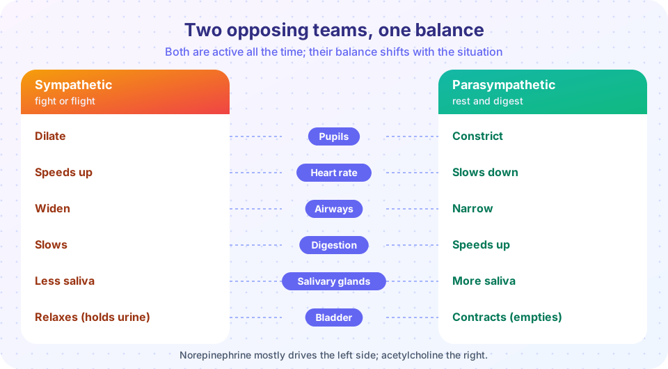
The sympathetic division prepares the body for activity and stress. Its preganglionic neurons start in the thoracic and upper lumbar spinal cord (T1 to L2), so it is called the thoracolumbar division. Its ganglia lie close to the spinal cord (the sympathetic chain), so preganglionic fibers are short and postganglionic fibers long. Acetylcholine (ACh) is the neurotransmitter at the ganglia and, for most targets, norepinephrine is released at the effector. (Sweat glands are a notable exception: they get ACh.) The adrenal medulla acts like a giant ganglion: when stimulated it releases mainly epinephrine (adrenaline) and some norepinephrine into the blood, which spreads and prolongs the response. Sympathetic effects are widespread because one preganglionic neuron synapses on many postganglionic neurons. The result: faster, stronger heartbeat, wider airways, dilated pupils, blood redirected to muscles, sweating, release of glucose from the liver, and slowed digestion.
Parasympathetic: rest and digest
The parasympathetic division conserves energy and supports digestion and recovery. Its neurons come from the brainstem (cranial nerves III, VII, IX and X) and the sacral spinal cord (S2 to S4), so it is called craniosacral. Its ganglia sit near or inside the target organ, so preganglionic fibers are long and postganglionic fibers short. ACh is the transmitter at both synapses. The vagus nerve (X) carries roughly three quarters of all parasympathetic fibers, supplying the heart, lungs and most digestive organs. Effects are more localized: slowing the heart, constricting pupils, narrowing airways, increasing digestive secretions and gut movement, and emptying the bladder.
| Feature | Sympathetic | Parasympathetic |
|---|---|---|
| Origin | thoracic and lumbar cord (T1 to L2) | brainstem (III, VII, IX, X) and sacral cord (S2 to S4) |
| Preganglionic fiber | short | long |
| Postganglionic fiber | long | short |
| Ganglia | near the spinal cord | near or in the target organ |
| Main effector transmitter | norepinephrine | acetylcholine |
| Spread of effect | widespread | localized |
| Overall role | mobilizes (stress, exercise) | restores (rest, digestion) |
Most organs receive both divisions, called dual innervation, and the two usually oppose each other, so the heart rate at any moment is the balance of the two. At rest, the parasympathetic influence on the heart dominates, which is why a resting heart rate of about 60 to 80 beats per minute is lower than the 100 or so beats per minute that the heart's own pacemaker would produce without that braking. Exceptions exist: most blood vessels have only sympathetic input, and sweat glands are sympathetic.
The enteric nervous system
The walls of the digestive tract contain their own network of neurons, the enteric nervous system, with hundreds of millions of nerve cells, which is more than the spinal cord. It can control gut movement and secretion on its own, and it is modulated by the sympathetic and parasympathetic systems. It is sometimes called the "second brain", although the phrase is a metaphor.
The slider below shows the idea of a balance between the two divisions. The numbers are an illustrative model of the direction of changes and of rough magnitudes, not physiological measurements.
Common misconceptions
- "Autonomic means automatic and cannot be influenced." Emotions, breathing techniques and training can change autonomic output, although not by direct will.
- "Sympathetic is on and parasympathetic is off, or the reverse." Both are active all the time; their balance shifts.
- "Reflexes happen in the brain." Many, such as the knee jerk, are processed entirely in the spinal cord.
- "The vagus nerve is only a calming nerve." It also carries sensory information from organs to the brain and has many functions.
Check your understanding
Quick check · 6 questions
of 6 answered You scored / 6 · perfect!
-
1. How many pairs of cranial nerves are there?
There are 12 pairs of cranial nerves and 31 pairs of spinal nerves. -
2. Which sequence correctly lists the parts of a reflex arc?
The signal flows from receptor to sensory neuron, then the CNS, then motor neuron, then effector. -
3. Which of these is a sympathetic effect?
Sympathetic activity widens the airways; the other effects are parasympathetic. -
4. Where do parasympathetic ganglia lie?
Parasympathetic ganglia are near the target, so postganglionic fibers are short. -
5. How many neurons does the autonomic pathway use between the CNS and the effector?
A preganglionic neuron synapses with a postganglionic neuron in a ganglion. -
6. Which spinal nerve plexus supplies the arm?
The brachial plexus (C5 to T1) supplies the upper limb.
Summary
- Nerves are bundles of axons wrapped in endoneurium, perineurium and epineurium; 12 pairs of cranial nerves and 31 pairs of spinal nerves link the CNS to the body.
- Spinal nerves form plexuses (cervical, brachial, lumbar, sacral); each supplies a dermatome.
- A reflex arc has five parts; the knee jerk is monosynaptic, and the withdrawal reflex is polysynaptic.
- The autonomic system uses a two-neuron pathway to control smooth muscle, cardiac muscle and glands.
- The sympathetic (thoracolumbar, norepinephrine) mobilizes; the parasympathetic (craniosacral, acetylcholine) restores; the enteric system governs the gut.
Next: The special senses
Track your progress
Create a free account to tick off lessons and pick up where you left off on any device.
of Anatomy and Physiology lessons completed