


















Lesson 19 of 52 · Nervous System and Senses
Neurons and nerve signals: action potentials and synapses
See how a neuron is built, how ion gradients set the resting potential, how an action potential fires and travels, and how synapses pass signals on.
A neuron is a cell that has evolved into a living wire. Its job is to pick up information at one end, carry it over a distance and hand it to the next cell. In your sciatic nerve a single axon can be more than a meter long, yet the cell body that supports it is a speck about 0.02 millimeters wide. The signal that travels along that axon is not a flow of electricity as in a copper cable. It is a self-renewing wave of changes in the voltage across the cell membrane, called an action potential, and it is built from the ion transport you studied in Chapter 2.
This lesson explains the neuron's structure, how the membrane holds a steady voltage at rest, how an action potential fires and moves, and how the signal crosses the gap to the next cell.
What you will learn
- The parts of a neuron and how myelin speeds conduction.
- How the sodium-potassium pump and leak channels set the resting potential.
- The phases of an action potential and why it is all-or-none.
- How chemical synapses work, and how signals are added up.
Key terms
- Resting membrane potential: the voltage across the membrane of an unstimulated neuron, about -70 mV.
- Action potential: a brief, all-or-none reversal of membrane voltage that travels along an axon.
- Threshold: the membrane voltage (about -55 mV) at which voltage-gated sodium channels open and an action potential begins.
- Synapse: the junction where one neuron passes a signal to another cell.
- Neurotransmitter: a chemical released at a synapse to carry the signal.
Structure of a neuron
A typical neuron has three regions.
- Dendrites are branching extensions that receive signals from other cells. Some neurons have a few; others have thousands of tiny spines on their dendrites.
- The cell body (soma) contains the nucleus and most organelles. It integrates the incoming signals.
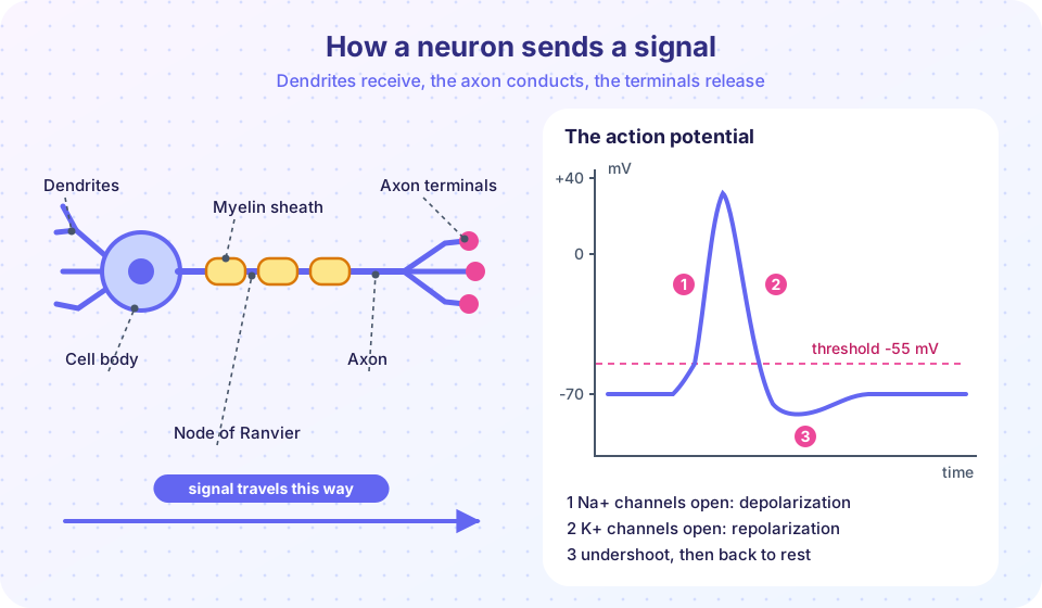
- The axon is a single long extension that carries the signal away. It starts at the axon hillock, the trigger zone where action potentials are generated, and ends in axon terminals (synaptic knobs) that release neurotransmitter.
Many axons are wrapped in myelin, a fatty insulating sheath made of glial cell membrane (oligodendrocytes in the CNS, Schwann cells in the PNS). The sheath is interrupted every millimeter or so by gaps called nodes of Ranvier. Myelin forces the electrical signal to jump from node to node, a process called saltatory conduction (from the Latin saltare, to leap), which raises the speed from about 1 m/s to as much as 100 m/s and saves energy. In multiple sclerosis, the immune system attacks myelin in the CNS, slowing or blocking signals.
By structure, neurons are multipolar (one axon, many dendrites; most neurons), bipolar (one dendrite, one axon; found in the retina) or unipolar (a single process that splits, as in most sensory neurons). By function they are sensory (afferent), motor (efferent) or interneurons (which connect other neurons inside the CNS, and make up the vast majority).
The resting membrane potential
Inside a resting neuron, the cell is about 70 millivolts more negative than the outside. That voltage difference, the resting membrane potential (RMP), arises from two facts.
First, ions are unevenly distributed. The Na⁺/K⁺ pump (Chapter 2) keeps sodium concentration high outside (about 145 mM) and low inside (about 15 mM), and potassium high inside (about 140 mM) and low outside (about 5 mM). Second, the resting membrane is far more permeable to K⁺ than to Na⁺, because it has many open potassium leak channels but few open sodium channels. Potassium leaks out down its gradient, leaving behind unbalanced negative charge, mainly from large negatively charged proteins that cannot leave. The outflow stops when the electrical pull back on K⁺ balances the chemical push out. That balance point is the equilibrium potential of the ion, given by the Nernst equation: E = (61.5 mV / z) × log10([ion]out / [ion]in) at body temperature (37 °C), where z is the ion's charge.
The calculator below computes equilibrium potentials for four ions using typical mammalian concentrations. The RMP of about -70 mV sits close to the potassium value (about -90 mV) but is pulled up a little by the small sodium leak. The numbers are typical textbook concentrations, which vary by cell.
Press Run (or Ctrl + Enter) to see the result here.
Program input · what you'd type while the program runs
Type the answers before you press Run, one per line. Each time the program reads a line (with input()), it gets the next line from this box.
Graded potentials and threshold
When a neuron receives input at its dendrites and soma, the membrane voltage changes by small amounts that spread a short distance and fade. These graded potentials vary in size with the strength of the stimulus. A change that makes the inside less negative is a depolarization and, if it is caused by excitatory input, is called an excitatory postsynaptic potential (EPSP). A change that makes it more negative is a hyperpolarization (an inhibitory postsynaptic potential, IPSP). The neuron adds up all the inputs: temporal summation (inputs in quick succession) and spatial summation (inputs from many places at once). If the sum at the axon hillock reaches about -55 mV, the threshold, an action potential fires. If not, nothing happens.
The action potential
Once threshold is reached, a stereotyped sequence takes place in about 2 milliseconds. It relies on two kinds of voltage-gated channels that open and close in response to the voltage.
- Depolarization. Voltage-gated Na⁺ channels open rapidly. Sodium floods in, and the membrane potential shoots up, reaching about +30 mV. Positive feedback drives this: more depolarization opens more channels.
- Repolarization. At about the peak, the Na⁺ channels inactivate (they close and cannot reopen for a while), and slower voltage-gated K⁺ channels open. Potassium rushes out, and the voltage falls back toward rest.
- Undershoot (hyperpolarization). The K⁺ channels close slowly, so the membrane briefly goes more negative than rest, about -80 mV, and then settles back at -70 mV as the Na⁺/K⁺ pump and leak channels restore the baseline.
Three rules follow. First, an action potential is all-or-none: a stimulus either reaches threshold and produces a full-size spike, or it does not produce one. A stronger stimulus makes more spikes per second, not bigger spikes. This is the nervous system's frequency code. Second, there is a refractory period: during the absolute refractory period (while Na⁺ channels are inactivated) no new spike is possible, and during the relative refractory period (the undershoot) a stronger stimulus is needed. This limits the firing rate to a few hundred per second and makes the spike move in only one direction. Third, only a tiny fraction of the ions move per spike, so a neuron can fire thousands of times before its gradients run down noticeably, but the pump needs a lot of ATP to keep pace.
Propagation. The spike at one patch of the axon spreads current to the next patch, which depolarizes to threshold and fires its own spike, and so on. In unmyelinated axons the wave moves continuously along the membrane. In myelinated axons the spike regenerates only at nodes of Ranvier, which is why conduction is faster. Larger-diameter axons also conduct faster.
Local anesthetics such as lidocaine work by blocking voltage-gated Na⁺ channels, so action potentials in pain fibers cannot be generated. It is a useful example of how understanding the channel explains a drug's effect, not advice about any treatment.
The following program is a leaky integrate-and-fire model. It is a deliberate simplification: instead of simulating channels, it treats the neuron as a leaky capacitor that charges toward a target voltage; when the voltage reaches -55 mV it records a spike (drawn as a vertical line to +30 mV) and resets to -70 mV. It shows how stronger input produces a higher firing rate, the frequency code.
Press Run (or Ctrl + Enter) to see the result here.
Program input · what you'd type while the program runs
Type the answers before you press Run, one per line. Each time the program reads a line (with input()), it gets the next line from this box.
Synapses: passing the signal on
A neuron does not touch the next cell. At a chemical synapse the axon terminal of the presynaptic neuron sits across a gap of about 20 nanometers, the synaptic cleft, from the postsynaptic cell. Transmission works as follows:
- The action potential arrives at the axon terminal and opens voltage-gated Ca²⁺ channels.
- Calcium entering the terminal makes synaptic vesicles fuse with the membrane and release neurotransmitter by exocytosis.
- The neurotransmitter diffuses across the cleft and binds to receptors on the postsynaptic membrane.
- The receptors open ion channels (or start internal signaling), producing an EPSP or IPSP.
- The signal ends as the neurotransmitter is removed: broken down by enzymes (acetylcholinesterase for acetylcholine), taken back up into the terminal (reuptake) or by nearby glia, or it diffuses away.
Synaptic transmission is one-way, and the delay across a synapse is only about 0.5 ms. Some neurons also connect through electrical synapses with gap junctions, which pass current directly and very fast (found in some reflex circuits and in heart muscle).
Whether a synapse excites or inhibits depends on the receptor, not on the chemical alone. Some of the most important transmitters are:
| Neurotransmitter | Main roles |
|---|---|
| Acetylcholine | neuromuscular junction, parasympathetic system, memory-related circuits |
| Glutamate | the main excitatory transmitter in the brain; learning |
| GABA | the main inhibitory transmitter in the brain |
| Dopamine | movement control, motivation and reward pathways |
| Serotonin | mood, sleep and appetite circuits |
| Norepinephrine | alertness; the sympathetic system |
| Endorphins | the body's natural pain-modulating peptides |
Many drugs and diseases act at synapses. In Parkinson's disease, dopamine-producing neurons in the substantia nigra die, causing movement problems. Many antidepressants change how serotonin is cleared from synapses, and alcohol and sedatives enhance GABA's inhibition. These are examples showing why synapses matter; they are not a guide to treatment, and the research on exactly how many drugs achieve their effects is still developing.
Common misconceptions
- "A stronger stimulus produces a bigger action potential." Spikes are all-or-none. Strength is encoded by the firing rate and the number of neurons active.
- "Nerve signals are electricity moving through the axon like in a wire." They are waves of voltage change driven by ions crossing the membrane, far slower than electrons in a wire.
- "Neurons cannot change." Synapses strengthen and weaken with use, which is the basis of learning, a property called plasticity.
- "Neurotransmitters are either excitatory or inhibitory by nature." The effect depends on the receptor on the postsynaptic cell.
Check your understanding
Quick check · 6 questions
of 6 answered You scored / 6 · perfect!
-
1. What mainly sets the resting membrane potential near -70 mV?
High membrane permeability to K+, together with ion gradients maintained by the pump, sets the resting potential. -
2. What happens first when a neuron reaches threshold?
Depolarization is caused by Na+ entry through voltage-gated sodium channels. -
3. How does the nervous system code a stronger stimulus?
Action potentials are all-or-none, so intensity is carried by how many spikes per second occur. -
4. Why is conduction faster in myelinated axons?
Myelin insulates the axon, so the spike regenerates only at nodes, which is saltatory conduction. -
5. Which event directly causes vesicles to fuse at the axon terminal?
Ca2+ entering through voltage-gated channels triggers exocytosis of neurotransmitter. -
6. What is the main inhibitory neurotransmitter of the brain?
GABA is the major inhibitory neurotransmitter, while glutamate is the major excitatory one.
Summary
- A neuron has dendrites (input), a cell body (integration) and an axon (output) that ends in terminals; myelin and nodes of Ranvier speed conduction.
- The resting potential of about -70 mV comes from ion gradients, K⁺ leak channels and the Na⁺/K⁺ pump.
- Inputs sum at the axon hillock; reaching about -55 mV fires an all-or-none action potential built from Na⁺ entry then K⁺ exit.
- Intensity is coded by firing rate; refractory periods make signals one-way and limit rate.
- At chemical synapses, calcium triggers vesicle release; transmitters bind receptors and are then cleared; different receptors determine excitation or inhibition.
Track your progress
Create a free account to tick off lessons and pick up where you left off on any device.
of Anatomy and Physiology lessons completed