


















Lesson 26 of 48 · Translation and Protein Folding
Protein Folding, Chaperones and Misfolding
Learn how a chain of amino acids folds into a working protein, from Anfinsen's experiment and the folding funnel to chaperones, misfolding diseases and AlphaFold.
A ribosome produces a chain. A protein is a chain that has folded into one particular shape. The difference matters, because the shape decides what the protein can do: which molecule an enzyme can grip, which pore a channel forms, which surface an antibody can recognize. A chain of 300 amino acids could in principle fold in an astronomical number of ways, yet inside a cell it finds the right one in milliseconds to minutes. This lesson explains how, and what goes wrong when it fails.
What you will learn
- The four levels of protein structure and which forces hold them.
- What Anfinsen's experiment showed and why Levinthal's paradox is not a real paradox.
- What chaperones do and why cells need them.
- How misfolding causes disease and how modern tools predict structures.
Key terms
Native state: the folded conformation in which a protein works.
Hydrophobic effect: the tendency of nonpolar side chains to cluster away from water, the main driving force of folding.
Chaperone: a protein that helps others fold or refold without being part of the final structure.
Denaturation: loss of the folded structure, for example by heat, extreme pH or urea.
Amyloid: insoluble fibrils made of stacked beta strands, linked to several diseases.
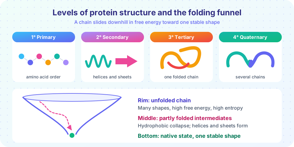
Four levels of structure
- Primary structure is the sequence of amino acids, set by the gene. Peptide bonds link the backbone.
- Secondary structure is the local pattern of the backbone, held by hydrogen bonds between backbone C=O and N–H groups. The two main patterns are the α-helix (3.6 residues per turn, with the C=O of residue i bonded to the N–H of residue i + 4, as Linus Pauling proposed in 1951) and the β-sheet (neighboring strands bonded side to side).
- Tertiary structure is the full three-dimensional shape of one chain. Hydrophobic collapse buries nonpolar side chains in the core, ionic and hydrogen bonds fine-tune the fit, and in secreted proteins disulfide bonds between cysteines add strong cross-links.
- Quaternary structure is the arrangement of several chains. Hemoglobin, for example, is a tetramer of two α and two β chains.
Many proteins also contain domains, compact units that fold independently and are reused in different proteins like words in a sentence, and some regions are intrinsically disordered and become ordered only when they bind a partner.
Why does a chain fold?
At room temperature water molecules form a dynamic network. A nonpolar side chain in water forces nearby water molecules into an ordered cage, which lowers the entropy of the water. When nonpolar groups gather in the protein core, that water is released, and the entropy of the whole system rises. This hydrophobic effect is the dominant driving force; hydrogen bonds, van der Waals contacts and salt bridges refine the result. The gain in stability of a typical protein is small, only about 20 to 60 kJ/mol net, which is a few hydrogen bonds' worth. That is why proteins are fragile and heat denatures them.
Anfinsen's experiment
In the early 1960s Christian Anfinsen studied ribonuclease A, a small enzyme with four disulfide bonds. He unfolded it with urea and a reducing agent and the enzyme lost all activity. When he removed the chemicals slowly, the enzyme folded again and regained activity. The conclusion, honored in the Nobel Prize in Chemistry in 1972, was that the amino acid sequence contains all the information needed to find the native structure. (There is a catch: if the disulfide bonds reform while the protein is still disordered, you get a scrambled enzyme, which shows that the order of events matters.)
Levinthal's paradox
Cyrus Levinthal pointed out in 1968 that a random search would take forever. Suppose each of 100 residues can take just 3 conformations. That gives 3^100, about 5 × 10^47 shapes. Even sampling 10^13 shapes per second, the search would take about 5 × 10^34 seconds, far longer than the age of the universe, about 4 × 10^17 seconds. Proteins do not search randomly. The energy landscape is shaped like a funnel: contacts that form early stabilize the chain and bias the search downhill. The paradox disappears once folding is seen as a guided process.
Helping the chain: chaperones
In the crowded cell, a half-folded chain exposes sticky hydrophobic patches that could cling to other chains and form aggregates. Chaperones prevent this.
- Trigger factor in bacteria sits at the ribosome exit tunnel and shields the chain as it emerges.
- Hsp70 (DnaK in E. coli) binds short hydrophobic stretches in an ATP-driven cycle, holding and releasing the chain so it can try again.
- Chaperonins such as GroEL/GroES in bacteria and TRiC in eukaryotes are barrel-shaped complexes. A chain goes inside the barrel, a lid closes, and the chain folds in isolation.
- Hsp90 helps mature many signaling proteins.
Chaperones do not tell a protein how to fold: Anfinsen's rule still holds. They increase the yield by preventing accidents. Many are heat-shock proteins because heat produces more unfolded chains.
In the endoplasmic reticulum, additional helpers such as protein disulfide isomerase fix wrong disulfide bonds and enzymes attach sugar chains. Many proteins are also trimmed after translation: the initiator methionine may be removed, signal peptides are cut off and chains can be phosphorylated, glycosylated or cleaved to become active.
When folding fails
If a protein cannot fold, the cell tags it with ubiquitin and sends it to the proteasome for destruction, a pathway recognized by the 2004 Nobel Prize in Chemistry (Aaron Ciechanover, Avram Hershko, Irwin Rose). When this quality control is overwhelmed or the mutation is severe, disease follows.
- In cystic fibrosis, the most common mutation deletes one phenylalanine (ΔF508) in the CFTR chloride channel; the protein misfolds and is degraded, so it never reaches the cell surface.
- In prion diseases a normal protein adopts an alternative, β-rich form that converts others (Stanley Prusiner, Nobel Prize 1997).
- In Alzheimer's and Parkinson's disease misfolded proteins accumulate as amyloid. Whether these aggregates cause the damage or mark it remains debated.
Predicting structure
For decades, determining a structure needed X-ray crystallography, NMR or cryo-electron microscopy. In 2020, DeepMind's AlphaFold 2 predicted structures at near-experimental accuracy for many proteins in the CASP14 competition, and public databases now hold predicted structures for hundreds of millions of sequences. Demis Hassabis and John Jumper shared the 2024 Nobel Prize in Chemistry with David Baker, who pioneered computational protein design. Predictions are not the same as folding pathways, and they are least reliable for disordered regions and for proteins that change shape, so experiments remain essential.
Common misconceptions
"A protein has only one shape." Many proteins flex or switch between states; the native state is an ensemble around a stable structure.
"Chaperones contain the folding instructions." They only prevent aggregation and give chains time to find the shape the sequence already encodes.
"Denaturation breaks peptide bonds." It disrupts weak interactions; the covalent peptide bonds stay intact.
Try it
The first example calculates Levinthal's numbers for different chain lengths and compares them with the age of the universe.
Press Run (or Ctrl + Enter) to see the result here.
Program input · what you'd type while the program runs
Type the answers before you press Run, one per line. Each time the program reads a line (with input()), it gets the next line from this box.
The second example draws a hydropathy plot of an invented sequence with the Kyte-Doolittle scale and a window of nine residues to keep the curve detailed (the original authors suggested a window of 19 residues and an average above about 1.6 to flag membrane-spanning helices). The sequence is a made-up teaching example.
Press Run (or Ctrl + Enter) to see the result here.
Program input · what you'd type while the program runs
Type the answers before you press Run, one per line. Each time the program reads a line (with input()), it gets the next line from this box.
The third example plots a helical wheel. An α-helix turns 100° per residue, so you can see whether one face of the helix is hydrophobic and the other polar (amphipathic). The sequence is a designed teaching peptide.
Press Run (or Ctrl + Enter) to see the result here.
Program input · what you'd type while the program runs
Type the answers before you press Run, one per line. Each time the program reads a line (with input()), it gets the next line from this box.
Check your understanding
Quick check · 5 questions
of 5 answered You scored / 5 · perfect!
-
1. What did Anfinsen's ribonuclease experiment show?
After the unfolding agents were removed, the enzyme refolded and became active again, so the sequence carries the information. -
2. Which force is usually the main driver of folding?
Burying nonpolar side chains releases ordered water, which raises entropy and favors the folded state. -
3. Levinthal's paradox is resolved by the idea that
Folding is biased by early stabilizing contacts, so the chain does not need to try every conformation. -
4. Which pair is matched correctly?
In an α-helix the backbone C=O of residue i bonds with the N-H of residue i + 4. -
5. In cystic fibrosis caused by ΔF508, the CFTR protein
The deletion of one phenylalanine causes misfolding, so quality control destroys most of the protein.
Summary
- Proteins have four levels of structure; the sequence determines the fold.
- The hydrophobic effect drives folding, and folding follows a funnel-shaped landscape.
- Chaperones prevent aggregation; they do not encode the shape.
- Misfolding underlies diseases such as cystic fibrosis, prion disease and amyloid disorders, and quality control degrades faulty proteins through the proteasome.
- AlphaFold-type tools predict structures well, though experiments are still needed.
Next: Chapter 5 review
Track your progress
Create a free account to tick off lessons and pick up where you left off on any device.
of Molecular Biology lessons completed