


















Lesson 43 of 48 · Projects and Practice
Case Study: Sickle-Cell Disease from Gene to Therapy
Follow one disease through the whole course: the HBB point mutation, the changed hemoglobin, diagnosis with restriction enzymes, population genetics and CRISPR therapy.
Few diseases show molecular biology as clearly as sickle-cell disease. One base in one gene is changed. One amino acid in one protein is different. A red blood cell changes shape, and a person suffers pain and organ damage. And, remarkably, the story ends with a therapy that edits the genome. In this case study you will use almost every idea from the course: the code, mutations, restriction enzymes, regulation of globin genes, population genetics, and CRISPR editing. As in a real analysis, you will move between sequence, protein, cell and patient.
This case study is educational information about science. It is not medical advice. Anyone with questions about sickle-cell disease or its treatment should speak with a health professional.
What you will learn
- How a single base change in HBB gives sickle hemoglobin and why the cells change shape.
- How the mutation was found and how restriction enzymes were used for diagnosis.
- Why the allele is common in some populations (heterozygote advantage).
- How CRISPR-based therapy works by switching fetal hemoglobin back on.
Key terms
Hemoglobin: the iron-containing protein of red blood cells that carries oxygen; adult HbA is two α and two β chains.
HbS: sickle hemoglobin, with valine instead of glutamate at position 6 of the β chain.
HbF: fetal hemoglobin, two α and two γ chains, made before birth and in small amounts afterwards.
Heterozygote advantage: higher fitness of carriers (Aa) than of either homozygote.
RFLP: restriction fragment length polymorphism, a difference in restriction fragments caused by a variant that creates or destroys a site.
From gene to protein
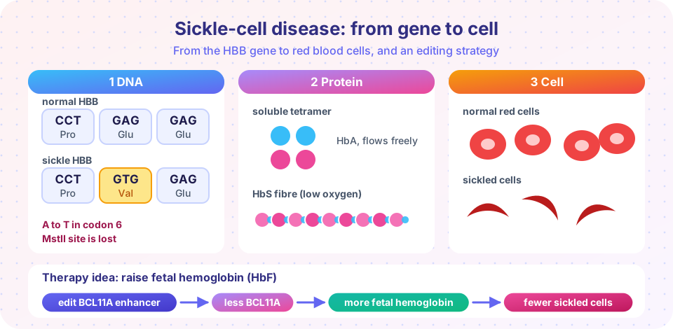
Adult hemoglobin consists of four chains. The β-globin chain is encoded by the HBB gene on chromosome 11, and its protein starts with the amino acids Val-His-Leu-Thr-Pro-Glu-Glu-Lys (the initiator methionine is removed). In the coding sequence these are the codons GTG CAC CTG ACT CCT GAG GAG AAG. In sickle-cell disease, the sixth amino acid of the mature chain, glutamate, is replaced by valine. The cause is a single base change in the DNA: the codon GAG (Glu) becomes GTG (Val), an A to T change, a missense mutation. In the notation you met earlier, it is called Glu6Val, or by the DNA change c.20A>T.
Linus Pauling and colleagues showed in 1949 that sickle hemoglobin has a different electrical charge from normal hemoglobin (it moves differently in an electric field), and they called sickle-cell anemia a "molecular disease", the first disease traced to a protein. In 1956 Vernon Ingram located the difference at one position by cutting the protein into peptides and comparing their patterns. Glutamate is negatively charged and hydrophilic; valine is neutral and hydrophobic.
Why does one amino acid matter?
When hemoglobin releases its oxygen, the β chains change shape and expose a hydrophobic pocket. In normal HbA the pocket is empty. In HbS, the valine sticking out at position 6 of one molecule fits into the pocket of a neighbor, so deoxygenated HbS molecules stack into long rigid fibers. The fibers distort the red blood cell into a crescent ("sickle") shape. Sickled cells are rigid and sticky: they clog small blood vessels (vaso-occlusion), causing episodes of severe pain and damaging organs, and they break down early, causing anemia. The effect is very sensitive to the concentration of HbS and to how long the cell stays deoxygenated, which is why cells in tissues with low oxygen suffer most, and why anything that lowers the HbS concentration, such as fetal hemoglobin, helps.
Inheritance and population genetics
The disease is autosomal recessive. A person who inherits the HbS allele from both parents (genotype SS) has the disease. Carriers (AS) have the sickle-cell trait and are usually healthy. Two carriers have a 1 in 4 chance of a child with the disease with every pregnancy.
Why is the allele common, since it is harmful in homozygotes? In regions where falciparum malaria is or was common (parts of sub-Saharan Africa, the Mediterranean, the Middle East and India), carriers are less likely to suffer severe malaria. So carriers (AS) have a higher average fitness than normal homozygotes (AA), who are more vulnerable to malaria, and than sufferers (SS). This heterozygote advantage (balancing selection) holds the allele at a stable frequency. With relative fitnesses AA = 1 − t, AS = 1 and SS = 1 − s, the equilibrium frequency of the S allele is q = t / (s + t). Most births with the disease today occur in sub-Saharan Africa, which makes access to testing and treatment a question of global health.
Diagnosis with a restriction enzyme
In the 1970s and 1980s the sickle mutation was one of the first genetic diseases diagnosed at the DNA level. Look at the sequence around codon 6: CCT GAG GAG. The normal sequence CCTGAGG fits the recognition site CCTNAGG of the restriction enzyme MstII (N is any base). The sickle mutation changes it to CCTGTGG, which no longer fits, so the site is lost. When DNA from a person is amplified or blotted and cut with MstII, the normal allele gives two smaller pieces and the sickle allele one larger piece. Yuet Wai Kan and colleagues used this for prenatal diagnosis. Today diagnosis relies on hemoglobin analysis (chromatography or electrophoresis) in newborn screening and on direct PCR and sequencing.
From biology to therapy
Several treatments exist. Hydroxyurea increases the amount of fetal hemoglobin and reduces crises; transfusions dilute HbS; a bone marrow transplant from a matched donor can cure the disease but needs a donor and carries risks.
The molecular insight behind new therapies is a lesson in regulation (chapter 6). Before birth, red cells make HbF (α₂γ₂). In the months after birth, the γ-globin genes are switched off and the β-globin gene is switched on. A key repressor of γ-globin is the transcription factor BCL11A. Babies with sickle-cell disease are protected in the first months of life by HbF, because HbF does not copolymerize well with HbS, and some adults who inherit mutations that keep γ-globin on (hereditary persistence of fetal hemoglobin) have much milder disease. This suggested a strategy: reactivate HbF.
In exa-cel (Casgevy), blood stem cells are collected from the patient, edited outside the body with CRISPR-Cas9 and returned after the patient's own marrow has been cleared by chemotherapy. The edit does not repair the sickle mutation. It disrupts an erythroid-specific enhancer of the BCL11A gene, so red cell precursors make less BCL11A (the rest of the body's BCL11A is untouched, since the enhancer acts only in red cell lineage cells) and therefore more γ-globin and HbF. The therapy was approved in the United Kingdom in November 2023 and in the United States in December 2023, along with a gene-addition therapy (lovo-cel, Lyfgenia) that uses a lentivirus to add a modified β-globin gene. In early trials, most treated patients were free of severe pain crises for at least a year, but the procedure is intensive, with a stay in hospital, and the price exceeds two million US dollars, so access is a serious equity problem.
Common misconceptions
"Sickle-cell disease is a disease of one ethnic group." The allele follows the geography of malaria, not race; it is also found around the Mediterranean, in the Middle East and in India, and many people of African ancestry do not carry it.
"Carriers have a mild form of the disease." Carriers are generally healthy; severe problems are rare and usually need extreme conditions.
"The CRISPR therapy repairs the sickle mutation." The approved therapy raises fetal hemoglobin by editing a different gene's enhancer; the HBB mutation remains.
Try it
The first example works on the HBB coding sequence: it translates the normal and sickle versions, shows the one-residue difference and searches both for the MstII site CCTNAGG.
Press Run (or Ctrl + Enter) to see the result here.
Program input · what you'd type while the program runs
Type the answers before you press Run, one per line. Each time the program reads a line (with input()), it gets the next line from this box.
The second example simulates heterozygote advantage. It starts with several different S allele frequencies and follows them for 300 generations with illustrative fitnesses (malaria costs the AA genotype 10 percent; the SS genotype loses 80 percent). They all converge to q = t / (s + t). The fitnesses are chosen for illustration and are not measurements of any population.
Press Run (or Ctrl + Enter) to see the result here.
Program input · what you'd type while the program runs
Type the answers before you press Run, one per line. Each time the program reads a line (with input()), it gets the next line from this box.
The third example is a toy model of why more fetal hemoglobin helps. Each cell has a random sensitivity; it sickles when the HbS concentration times the deoxygenation passes a steep threshold. It is a cartoon, not a physiological model.
Discussion and mini-project
Use the course to write a one-page analysis, with these questions as an outline.
- Which codon changes, and what kind of mutation is it? Why does valine matter more than another change, say Glu to Asp?
- How does the loss of an MstII site allow DNA diagnosis? Which fragment sizes would you expect after a PCR of the region?
- If 1 in 100 newborns in a region has the disease, what is the carrier frequency (use Hardy-Weinberg)?
- Why does raising HbF help, and why edit an enhancer rather than the gene itself?
- What are the benefits, risks and ethical questions of a therapy that costs millions and needs a hospital stay?
Check your understanding
Quick check · 5 questions
of 5 answered You scored / 5 · perfect!
-
1. What is the molecular change in sickle-cell disease?
A single A to T change in codon 6 of the mature chain replaces glutamate with valine. -
2. Why is the MstII site lost in the sickle allele?
The A in the fifth position of the site becomes T, so the recognition sequence is destroyed. -
3. Why does the HbS allele stay common in malaria regions?
Heterozygote advantage: carriers resist severe malaria and do not have the disease. -
4. What does the exa-cel therapy edit?
Reducing BCL11A in red cell precursors releases γ-globin, which makes HbF. -
5. Two carriers have a child. What is the chance that the child has sickle-cell disease?
Each parent passes the S allele with probability 1/2: 1/2 × 1/2 = 1/4.
Summary
- Sickle-cell disease results from a single missense mutation (Glu6Val) in HBB; deoxygenated HbS polymerizes and sickles red cells.
- The mutation destroys an MstII site, which allowed DNA-level diagnosis.
- The allele is common where malaria was common, because carriers have a fitness advantage.
- Fetal hemoglobin protects, and CRISPR editing of the BCL11A enhancer reactivates it.
- Access, cost and ethics are as important as the molecular biology.
Track your progress
Create a free account to tick off lessons and pick up where you left off on any device.
of Molecular Biology lessons completed