


















Lesson 5 of 48 · Getting Started
The Toolkit: Model Organisms, PCR, Gels and Sequencing
Meet the core methods of molecular biology: model organisms, PCR amplification, gel electrophoresis and DNA sequencing, with calculators that show how each works.
Molecular biology moves forward when someone invents a way to see or change molecules. A handful of techniques appear in almost every lab, and understanding them will make the rest of this course concrete. This lesson introduces the organisms researchers use, and three methods you will meet repeatedly: the polymerase chain reaction (PCR), gel electrophoresis and DNA sequencing.
Later chapters add cloning, CRISPR and genomics. Here the goal is the logic: what each method asks of a DNA sample and what it returns.
What you will learn
- Why a few model organisms support so much of molecular biology.
- How PCR makes billions of copies of one chosen DNA segment.
- How gel electrophoresis separates DNA fragments by size.
- How Sanger and next-generation sequencing read the order of bases.
Key terms
Model organism: a species studied intensively because it is easy to grow, breed and manipulate.
Primer: a short DNA oligonucleotide that anneals to a template and gives DNA polymerase a starting point.
Amplicon: the DNA segment copied by PCR.
Electrophoresis: moving charged molecules through a medium with an electric field.
Read: one sequence obtained from a sequencing machine.
Model organisms
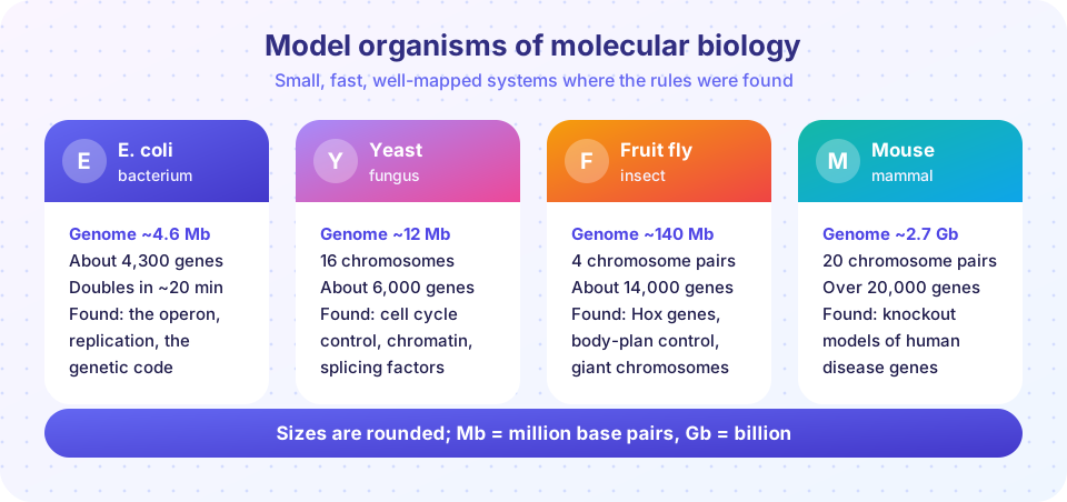
The cards above give rounded numbers for four classics. E. coli divides about every 20 minutes in rich medium, so a colony grows overnight; it taught us operons, replication and the genetic code. Baker's yeast is a single-celled eukaryote that is as easy to grow as a bacterium; it taught us the cell cycle and gene regulation in cells with a nucleus. The fruit fly Drosophila melanogaster showed how genes pattern a body, including the Hox genes that set the order of segments. The mouse is a mammal whose genes can be switched off one at a time to model human disease.
Other models fill other gaps: the roundworm C. elegans (every cell lineage mapped), zebrafish (transparent embryos), the plant Arabidopsis and the African clawed frog. They are good starting points because their basic machinery is conserved. A discovery in yeast is often valid for human cells.
PCR: photocopying one segment
Kary Mullis conceived PCR in 1983, and the first paper appeared in 1985. A PCR mix contains the template DNA, two primers that flank the target, a heat-stable DNA polymerase (from the hot-spring bacterium Thermus aquaticus, called Taq), the four nucleotides (dNTPs) and buffer. The mix is cycled through three temperatures:
- Denature at about 95 °C: the double strands separate.
- Anneal at about 50 to 65 °C: primers bind their complementary sites.
- Extend at 72 °C: the polymerase builds a new strand from each primer, 5′ to 3′.
Each cycle doubles the number of target molecules, so after n cycles an ideal reaction has 2ⁿ copies per starting copy. Thirty cycles give about 10⁹, a billion-fold amplification, in a couple of hours. Real efficiency is a little below 100 percent per cycle, and the reaction eventually plateaus when reagents run out.
Primer design matters. A primer is usually 18 to 25 nucleotides long, has a balanced GC content (about 40 to 60 percent) and a melting temperature (Tm) matched to its partner. A rough rule, the Wallace rule, estimates Tm = 2 × (A + T) + 4 × (G + C) degrees Celsius for short oligos.
Gel electrophoresis: sorting by size
DNA carries a negative charge on every phosphate, so in an electric field it travels toward the positive electrode. In a porous gel of agarose or polyacrylamide, long fragments are slowed more than short ones, so smaller fragments run farther. Over a useful range, the distance traveled is roughly linear in the logarithm of fragment length. A lane of fragments of known sizes (a ladder) lets you read off the sizes of unknown bands by comparison. A dye such as ethidium bromide or a safer alternative lights up the DNA under UV or blue light.
Sequencing: reading the letters
In 1977 Frederick Sanger and colleagues introduced chain-termination sequencing. A polymerase copies a template in the presence of normal nucleotides plus a small fraction of dideoxy nucleotides (ddNTPs) that lack the 3′-OH group needed to extend the chain. Each time a ddNTP is incorporated, the chain stops. A set of fragments ending at every possible position is produced, separated by size, and the terminal bases are read in order. Modern capillary instruments use four fluorescent dyes and read up to about 800 to 1000 bases at a time.
Next-generation sequencing (NGS) changed the scale. Platforms such as Illumina read millions to billions of short fragments (typically 50 to 300 bases) in parallel. Long-read technologies (PacBio and Oxford Nanopore) read tens of thousands of bases or more from single molecules, which helps assemble repetitive regions. Chapter 8 covers how short reads are assembled into genomes.
| Method | What it gives | Typical strength |
|---|---|---|
| PCR | Many copies of one segment | Sensitivity and speed |
| Gel electrophoresis | Fragment sizes | Cheap size check |
| Sanger sequencing | Sequence of one fragment | Accuracy for one target |
| Short-read NGS | Billions of short reads | Whole genomes, expression |
| Long-read sequencing | Reads of kilobases to megabases | Structure and repeats |
Common misconceptions
"PCR copies the whole genome." It copies only the segment between the two primers. Ten starting copies become about ten billion copies of the same short target.
"A smear on a gel means the experiment worked." Sharp bands at expected sizes are the signal; a smear can mean degraded DNA or too much sample.
"Sequencing tells you what genes do." It tells you the order of bases. Function needs additional experiments.
Try it
First, a PCR calculator. It shows how the number of copies grows when efficiency is 100 percent and when it is lower, and where a plateau stops growth. The plateau is a simple model, not a measured curve.
Press Run (or Ctrl + Enter) to see the result here.
Program input · what you'd type while the program runs
Type the answers before you press Run, one per line. Each time the program reads a line (with input()), it gets the next line from this box.
Second, a virtual gel. Each fragment moves a distance that falls linearly with the logarithm of its length. This is a simplified model; real gels also depend on gel percentage, voltage and run time.
Press Run (or Ctrl + Enter) to see the result here.
Program input · what you'd type while the program runs
Type the answers before you press Run, one per line. Each time the program reads a line (with input()), it gets the next line from this box.
Check your understanding
Quick check · 5 questions
of 5 answered You scored / 5 · perfect!
-
1. After 10 ideal PCR cycles, how many copies come from one template molecule?
Each cycle doubles the number: 2^10 = 1,024. -
2. Which temperature step lets the primers bind?
At the annealing temperature the primers pair with their complementary sites on the single-stranded template. -
3. In an agarose gel, which DNA fragment travels farthest?
Smaller fragments move through the pores more easily, so they migrate farther. -
4. What makes Sanger sequencing stop a growing DNA chain?
A ddNTP can be added but then no further nucleotide can be attached, so the chain terminates. -
5. Why is a heat-stable polymerase used in PCR?
Taq polymerase is not destroyed by the high temperature of each denaturation step.
Summary
- Model organisms are small, fast and well characterized, so discoveries there often apply broadly.
- PCR amplifies the segment between two primers by cycling denature, anneal and extend; ideal copies = 2ⁿ per template.
- Gel electrophoresis sorts DNA by size; migration is roughly linear in log(size).
- Sanger sequencing uses chain terminators; next-generation sequencing reads millions of fragments in parallel.
Next: Chapter 1 Review
Track your progress
Create a free account to tick off lessons and pick up where you left off on any device.
of Molecular Biology lessons completed