


















Lesson 10 of 48 · The Molecules of Life
Amino Acids and the Four Levels of Protein Structure
Learn the 20 amino acids, how the peptide bond joins them and how a sequence folds into helices, sheets and 3D shapes, with hydropathy and peptide-mass calculators.
Proteins carry out most of the work of the cell: enzymes speed up reactions, structural proteins give cells their shape, antibodies defend the body and transport proteins move molecules across membranes. A protein is a chain of amino acids, yet the same kind of chain can be an enzyme, a muscle fiber or a hormone. The secret is how the chain folds.
This lesson introduces the 20 standard amino acids, the peptide bond that links them and the four levels of structure that turn a linear sequence into a working three-dimensional machine.
What you will learn
- The common structure of an amino acid and the four classes of side chains.
- How a peptide bond forms and why a chain has an N-terminus and a C-terminus.
- Primary, secondary, tertiary and quaternary structure, and the bonds behind each.
- Why one amino acid change can cause a disease such as sickle cell anemia.
Key terms
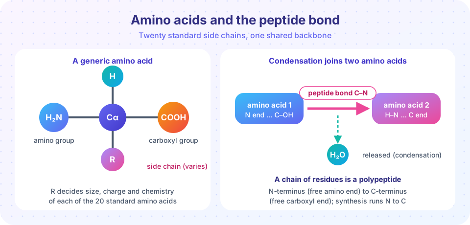
Amino acid: a molecule with an amino group, a carboxyl group, a hydrogen and a variable side chain on one central carbon.
Residue: one amino acid unit inside a chain.
Polypeptide: a chain of amino acids joined by peptide bonds.
α-helix and β-sheet: the two most common local folding patterns, held by hydrogen bonds in the backbone.
Hydrophobic effect: the tendency of oily groups to cluster away from water.
Amino acids and their side chains
Every standard amino acid has a central carbon (the α-carbon) bonded to an amino group (−NH₂), a carboxyl group (−COOH), a hydrogen atom and a side chain called R. In proteins the amino acids are of the L form. What differs is R, which sets size, charge and chemistry. The 20 standard amino acids fall into four broad classes:
| Class | Amino acids (one-letter code) | Behavior |
|---|---|---|
| Nonpolar (hydrophobic) | G, A, V, L, I, M, P, F, W | Cluster inside the protein, away from water |
| Polar, uncharged | S, T, C, Y, N, Q | Form hydrogen bonds with water and each other |
| Acidic (negative) | D, E | Carry a negative charge at cell pH |
| Basic (positive) | K, R, H | Carry a positive charge (histidine partly) |
A few have special roles. Glycine is the smallest, with only a hydrogen as its side chain, so it fits into tight turns. Proline's side chain loops back to its own backbone nitrogen, which makes kinks. Cysteine contains a sulfur and two cysteines can form a covalent disulfide bridge. Two rare additions, selenocysteine and pyrrolysine, are encoded in a few organisms by special use of stop codons.
The peptide bond
When the carboxyl group of one amino acid reacts with the amino group of the next, a molecule of water is lost and a peptide bond (a C-N bond) forms. This is a condensation reaction, like the formation of the phosphodiester bond in nucleic acids. The peptide bond has partial double-bond character, so the six atoms around it lie in a flat plane and can't rotate freely. Only the bonds on either side of each α-carbon rotate, and the angles allowed (called phi and psi) limit the shapes the backbone can adopt. The plot of allowed angles is a Ramachandran plot.
A chain has a free amino group at one end, the N-terminus, and a free carboxyl group at the other, the C-terminus. The ribosome builds proteins from N to C, and a sequence is written in that order. The average mass of an amino acid residue is about 110 daltons (Da), so a protein of 300 residues weighs roughly 33 kDa.
Four levels of structure
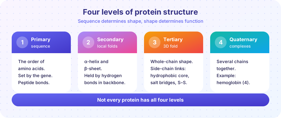
Primary structure is the amino acid sequence, specified by the gene. It is the only level encoded directly.
Secondary structure is local folding of the backbone, stabilized by hydrogen bonds between backbone C=O and N-H groups. Linus Pauling predicted the two main patterns in 1951. In an α-helix, the chain coils with 3.6 residues per turn and a pitch of about 0.54 nm, with each C=O bonding the N-H four residues ahead. In a β-sheet, strands lie side by side, with hydrogen bonds between neighboring strands; they can run in the same direction (parallel) or opposite (antiparallel).
Tertiary structure is the overall 3D shape of one chain. It arises from the hydrophobic effect (oily side chains pack into a core), hydrogen bonds, ionic attractions between oppositely charged side chains, and disulfide bridges. Tertiary structure creates binding sites and active sites with exact geometry.
Quaternary structure is the arrangement of several chains (subunits) into one complex. Hemoglobin, for example, has four subunits (two α and two β) that cooperate to carry oxygen. Not all proteins have this level: many are single chains.
Sequence determines structure
In the 1950s and 1960s Christian Anfinsen showed that the enzyme ribonuclease A, unfolded by urea and a reducing agent, spontaneously refolded and regained activity once the chemicals were removed. The information for the fold is in the sequence. This work earned him a share of the 1972 Nobel Prize. Folding in cells is often helped by chaperones (chapter 5), and predicting the fold from sequence was a famous challenge solved to a large extent by AI programs such as AlphaFold in 2020 and 2021.
The link between sequence and function is vivid in sickle cell anemia. A single DNA change turns the sixth amino acid of the β-globin chain from glutamic acid (acidic, charged) to valine (nonpolar). The new oily patch makes hemoglobin molecules stick to each other when oxygen is low, bending red cells into a sickle shape.
Common misconceptions
"A protein is a rigid object." Proteins move and flex, and many have flexible regions that are essential to function.
"Every protein has all four levels." Every protein has primary, secondary and tertiary structure in some form, but quaternary structure exists only where several chains assemble.
"Peptide bonds are formed by hydrogen." They are covalent C-N bonds made by condensation; hydrogen bonds stabilize folds but do not link the chain.
Try it
A hydropathy plot shows where a protein is oily or watery. Each amino acid has a Kyte-Doolittle value (positive means hydrophobic). A sliding-window average along the sequence reveals stretches rich in hydrophobic residues, which often span a membrane. The sequence below is invented for demonstration.
Press Run (or Ctrl + Enter) to see the result here.
Program input · what you'd type while the program runs
Type the answers before you press Run, one per line. Each time the program reads a line (with input()), it gets the next line from this box.
Second, a peptide-mass calculator. It sums average residue masses and adds one water for the free ends. The values are standard average masses in daltons.
Press Run (or Ctrl + Enter) to see the result here.
Program input · what you'd type while the program runs
Type the answers before you press Run, one per line. Each time the program reads a line (with input()), it gets the next line from this box.
Check your understanding
Quick check · 5 questions
of 5 answered You scored / 5 · perfect!
-
1. Which part of an amino acid differs among the 20 standard amino acids?
All share the same backbone; the side chain R makes each one unique. -
2. What is released when a peptide bond forms?
Peptide bond formation is a condensation reaction that removes one water molecule. -
3. Which level of structure is directly encoded by the gene?
The gene specifies the amino acid sequence; the other levels follow from it. -
4. Which interaction mainly stabilizes an alpha helix?
Each backbone C=O bonds to an N-H four residues along the chain. -
5. Sickle cell anemia results from replacing glutamic acid with
The Glu6Val change creates a hydrophobic patch that makes hemoglobin aggregate.
Summary
- Amino acids share a backbone and differ in their side chains, which are nonpolar, polar, acidic or basic.
- A peptide bond joins amino acids by condensation; chains run from N-terminus to C-terminus.
- Primary structure is the sequence; secondary is helices and sheets; tertiary is the 3D fold; quaternary is a complex of chains.
- The sequence determines the fold (Anfinsen), so one residue change can alter function, as in sickle cell anemia.
Track your progress
Create a free account to tick off lessons and pick up where you left off on any device.
of Molecular Biology lessons completed