


















Lesson 35 of 48 · Recombinant DNA and CRISPR
Gel Electrophoresis, Blotting and DNA Fingerprints
Learn how agarose gels separate DNA by size, how to estimate fragment lengths from a ladder, and how Southern blots and STR profiles identify sequences and people.
DNA is invisible and tiny, so how do you know whether your digest worked, whether your PCR gave one product or three, or whether two samples came from the same person? The standard answer is gel electrophoresis. You load a sample into a slab of gel, apply an electric field and watch fragments sort themselves by size. It is the single most-used readout in a molecular biology lab, and the reasoning that goes with it is the part worth learning: reading a ladder, spotting a failed reaction, and estimating a size from a picture.
What you will learn
- Why DNA moves in an electric field and why small fragments move farther.
- How to estimate a fragment size from a ladder using a log plot.
- Why plasmid DNA shows several bands.
- What Southern blots and STR DNA profiles reveal.
Key terms
Agarose gel: a porous gel made from seaweed polysaccharide, used to separate DNA fragments from about 100 bp to tens of kilobases.
DNA ladder (marker): a mix of fragments of known sizes run beside the samples.
Supercoiled plasmid: a circular DNA that is twisted on itself and runs faster than the same plasmid in other forms.
Southern blot: transfer of DNA fragments from a gel to a membrane, followed by detection with a labeled probe.
STR: short tandem repeat, a microsatellite whose number of repeats varies between people.
How a gel works
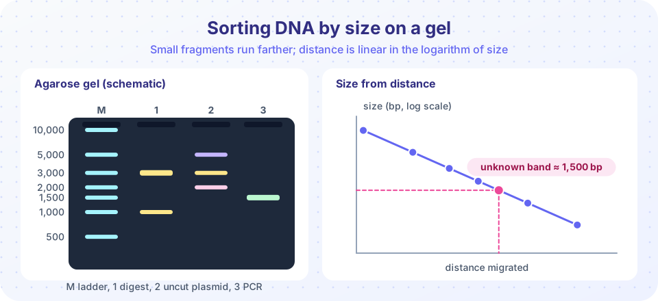
DNA has a negatively charged phosphate backbone, so in an electric field it moves toward the positive electrode (anode). The gel acts as a sieve: a fragment must wriggle through a mesh of pores. Small fragments slip through easily and move far; large fragments are held back. Because every nucleotide carries about the same charge, all fragments have nearly the same charge per unit of length, and the size of the pores, not charge, makes the separation.
A helpful rule: the distance migrated is roughly linear in the logarithm of the fragment size over a useful range. So the ladder is plotted on a semi-log graph, the points form a line, and you can read off the size of an unknown band from its distance. Low-percentage agarose (0.7 percent) separates large fragments (about 1 to 10 kb); higher percentages (2 percent) separate small ones. For fragments only a few to a few hundred bases long, polyacrylamide gels give resolution down to a single nucleotide.
To see the DNA you stain it with a dye that fluoresces when it binds DNA: ethidium bromide (a mutagen, handled with care) or safer dyes such as SYBR Safe. The gel is photographed under UV or blue light.
Reading a gel
When you look at a picture, ask four questions.
- Is the ladder clear? If not, the run or the staining failed.
- What is the size of each band? Use the ladder.
- Is the pattern what you predicted? Compare to a digest or PCR prediction (you can compute the expected sizes from the sequence).
- Do the controls behave? A negative control with no template should show no band.
Plasmids often give more than one band even when pure, because the same molecule can exist in different shapes. Supercoiled circles are compact and run fastest, linear molecules run slower, and nicked ("open circular") molecules run slowest. Cutting with a restriction enzyme converts all forms into one linear form, so digests give cleaner bands. This is one reason a diagnostic digest is used to check clones.
Bands also vary in brightness: the amount of fluorescent dye bound is proportional to the amount of DNA, so a thick band of a 3,000 bp fragment looks brighter than a band of the same molar amount of a 500 bp fragment (which has fewer base pairs to bind dye).
Blotting: finding a specific sequence
A gel shows all fragments, but you may want to know which one contains a particular gene. In 1975 Edwin Southern invented a solution. After electrophoresis, the DNA in the gel is denatured and transferred to a membrane by capillary action, keeping the pattern. The membrane is incubated with a labeled single-stranded probe that pairs with the sequence of interest, and washed, and only fragments that match light up. This is the Southern blot. By analogy, the Northern blot detects RNA, and the Western blot detects proteins using antibodies (after protein separation by SDS-PAGE). Blots are slower than PCR and have been replaced for many uses, but they remain valuable for checking the structure of an integrated transgene.
DNA fingerprinting
In 1984 Alec Jeffreys found regions of the human genome that differ greatly between people because they contain repeated units repeated a variable number of times. Modern forensic profiles use short tandem repeats (STRs): stretches such as GATA repeated 8 to 20 times. A PCR with fluorescent primers amplifies a set of STR loci (the US CODIS system uses 20 core loci), and electrophoresis (today in capillaries) sizes the products. Each person carries two alleles per locus, one from each parent. The combination across many loci is practically unique, except for identical twins. Matching probabilities are computed from the allele frequencies in the population, using the product rule for independent loci. The same technique is used to test parentage, since a child's alleles must come from the parents.
Common misconceptions
"DNA moves because it is attracted to the gel." It moves because of the electric field; the gel only slows it by size.
"Big fragments move farther because they are heavier." The opposite: small fragments move farther because they pass through the pores more easily.
"One band always means one product." Two different fragments of nearly the same size can run as a single band, which is why digest and sequencing confirm important results.
Try it
The first example fits the semi-log relationship to a ladder and estimates the sizes of unknown bands from their measured distances. The distances here are computed from the ideal model plus a small error, so they are teaching data and not a measurement.
Press Run (or Ctrl + Enter) to see the result here.
Program input · what you'd type while the program runs
Type the answers before you press Run, one per line. Each time the program reads a line (with input()), it gets the next line from this box.
The second example draws a simulated gel from a list of fragment sizes in several lanes. It is a rendering of an idealized model, not a photo.
Press Run (or Ctrl + Enter) to see the result here.
Program input · what you'd type while the program runs
Type the answers before you press Run, one per line. Each time the program reads a line (with input()), it gets the next line from this box.
The third example is an interactive run: slide the run time and watch fragments migrate. The model (distance proportional to time and decreasing with the logarithm of size) is a teaching simplification.
Check your understanding
Quick check · 5 questions
of 5 answered You scored / 5 · perfect!
-
1. In which direction does DNA move in an agarose gel?
The phosphate backbone is negatively charged, so DNA migrates toward the anode (+). -
2. Which fragment travels the farthest?
Smaller fragments move through the pores more easily. -
3. A plot of distance migrated against the log of fragment size for a ladder is
Over a useful range, distance is linear in log size, so you can read an unknown size from the line. -
4. What does a Southern blot detect?
DNA fragments are transferred to a membrane and the sequence of interest is found by hybridization. -
5. Why are STRs useful for identification?
Repeat counts vary, and combining many loci gives a highly specific profile.
Summary
- DNA is negatively charged and moves toward the positive electrode; the gel sorts fragments by size, smaller ones farther.
- Distance is linear in log size, so a ladder gives sizes of unknown bands.
- Plasmids show several bands (supercoiled, linear, nicked); digests simplify them.
- Southern blots find a sequence among fragments; STR profiles identify individuals by repeat counts.
- Always check ladders and controls before interpreting a gel.
Track your progress
Create a free account to tick off lessons and pick up where you left off on any device.
of Molecular Biology lessons completed