


















Lesson 18 of 46 · Hemodynamics and Genetic Disease
Thrombosis, Embolism, Infarction and Shock
Learn Virchow's triad, how thrombi form and what happens to them, the types of embolism, white versus red infarcts, and the main forms of shock.
Blood must flow freely, and it must also clot instantly when a vessel is cut. The same chemistry does both, which means that it can go wrong in either direction. When blood clots inside an intact vessel it forms a thrombus. If that thrombus, or any other mass, breaks free and lodges downstream it is an embolus. If the supply to a tissue is cut off long enough, the tissue dies: an infarct. In the extreme, when the whole circulation cannot supply the body, the result is shock. These are among the most common causes of death in the developed world, and they all start from the same few principles.
What you will learn
- The three elements of Virchow's triad with examples of each.
- What a thrombus looks like and what can happen to it.
- The main types of embolism, especially pulmonary embolism.
- How infarcts differ (white or red) and what determines the outcome.
- The classes of shock and its stages.
Key terms
Thrombus: a solid mass of platelets, fibrin and cells formed in a living vessel or heart chamber.
Embolus: a detached mass carried in the blood to a distant site.
Infarct: an area of ischemic necrosis caused by loss of arterial supply or venous drainage.
Ischemia: reduced blood supply to a tissue.
Shock: a state of generalized hypoperfusion of tissues.
Thrombosis and Virchow's triad
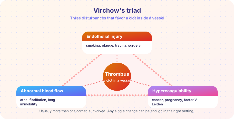
In 1856 Rudolf Virchow described three factors that favor a clot in a vessel. They are still called Virchow's triad.
- Endothelial injury. The lining of a healthy vessel actively prevents clotting. When damaged, it exposes collagen and tissue factor and loses its anticlotting properties. Causes: atherosclerotic plaques, the inner wall of the heart after a heart attack, trauma, surgery, smoking, and high blood pressure.
- Abnormal blood flow. Normal laminar flow keeps platelets in the center and washes activated clotting factors away. Stasis (slow or stagnant flow) occurs in atrial fibrillation, in a dilated aneurysm, in long periods of immobility (such as a long flight or bed rest) and in thickened blood (polycythemia). Turbulence damages the wall and creates eddies.
- Hypercoagulability. The blood itself clots too easily. Inherited causes include factor V Leiden, a common mutation that makes factor V resistant to inactivation by protein C, and a prothrombin gene mutation; deficiencies of antithrombin, protein C or protein S are rarer. Acquired causes include cancer, pregnancy, estrogen-containing hormone therapy, major surgery, severe trauma and some autoimmune conditions.
Often several elements combine. A patient confined to bed after a major operation has stasis (immobility), endothelial injury (surgery) and hypercoagulability (the stress response).
What a thrombus looks like
Thrombi can form in arteries, veins, heart chambers and capillaries. Arterial thrombi are often rich in platelets and usually form where flow is fast and the wall is injured. Venous thrombi form in slow flow and are rich in red cells and fibrin (red thrombi). A thrombus formed in life often shows lines of Zahn, alternating pale layers (platelets and fibrin) and dark layers (red cells), because it was built up in flowing blood. A post-mortem clot is a smooth, jelly-like mass without layers and is not attached to the wall.
Possible fates
- Propagation: the thrombus grows and may block the vessel.
- Embolization: pieces break off and travel.
- Dissolution: plasmin breaks it down, particularly in young thrombi.
- Organization and recanalization: the thrombus is invaded by inflammatory cells and fibroblasts and becomes fibrous; channels may form through it and restore some flow.
Deep vein thrombosis
Thrombi in the deep veins of the legs (deep vein thrombosis, DVT) cause swelling and pain in the leg, though many are silent. They are dangerous because they are the main source of pulmonary emboli. Superficial vein thrombi are painful but rarely embolize.
Embolism
An embolus is any solid, liquid or gas mass that travels in the blood to a site far from where it started. About 99 percent are fragments of thrombi (thromboembolism). The place where the embolus lodges depends on where it started.
| Type | Source | Typical consequence |
|---|---|---|
| Pulmonary thromboembolism | Deep leg veins (right heart and lungs) | Breathlessness, chest pain, strain on the right heart; a large embolus lodged at the main pulmonary artery split (saddle embolus) can cause sudden death |
| Systemic thromboembolism | Left side of the heart, from atrial fibrillation or a heart attack's mural clot | Stroke, kidney, spleen or leg artery blockage |
| Fat embolism | Fat released after fracture of long bones | Breathing difficulty, confusion and pinpoint rash a day or two after injury |
| Air embolism | Air entering veins (surgery, trauma), or nitrogen bubbles in decompression sickness | Obstruction of small vessels; the "bends" in divers |
| Amniotic fluid embolism | Fetal material entering the mother's blood in labor | Rare, severe obstetric emergency |
| Tumor embolism | Pieces of tumor | Metastasis |
In a paradoxical embolism, a venous embolus crosses from right to left through a hole in the heart (such as a patent foramen ovale) and reaches an artery.
Pulmonary embolism deserves attention. Most are small and cause no symptoms. The lung has a dual blood supply (pulmonary arteries and bronchial arteries), so even if a small branch is blocked, an infarct is uncommon unless the lungs or heart are already diseased. A large embolus suddenly raises the pressure in the right heart, which may fail.
Infarction
An infarct is an area of ischemic necrosis. The most common cause is arterial occlusion by thrombosis or embolism. Other causes include vasospasm, a plaque that suddenly enlarges, compression of a vessel by a tumor, and twisting of an organ on its stalk (torsion of the testis, twisting of the bowel).
White and red infarcts
- White (anemic) infarcts occur with arterial blockage in solid organs with end-arterial circulation, such as the heart, spleen and kidney. The tissue is pale and firm, and the infarct is wedge-shaped, with the blocked vessel at the point of the wedge.
- Red (hemorrhagic) infarcts occur with venous blockage (for example twisting of an organ) or in loose tissues like the lung, where blood leaks into the dead tissue, or in tissue with dual supply or previous congestion, such as the bowel.
Over days the infarct goes through a sequence like that of necrosis and repair: coagulative necrosis (liquefactive in the brain), an inflammatory border, then granulation tissue and finally a scar. A heart attack follows this pattern, which is why a heart infarct is softest, and at greatest risk of rupture, roughly between the third and seventh days.
What determines the outcome?
- The nature of the blood supply. Organs with two supplies (lung, liver) or rich collaterals (hand, forearm) tolerate blockage better.
- The speed of occlusion. Slow blockage lets collaterals develop.
- The vulnerability of the tissue. Neurons die after a few minutes; heart muscle after tens of minutes; fibroblasts survive hours.
- Oxygen content of the blood. Anemia or low oxygen make infarction more likely.
Shock
Shock is a state in which the circulation cannot deliver enough blood to tissues. The main types:
- Cardiogenic shock: the heart pump fails (large heart attack, arrhythmias, pulmonary embolism).
- Hypovolemic shock: too little blood volume (hemorrhage, severe vomiting or diarrhea, burns).
- Septic shock: severe infection triggers widespread inflammatory mediators (TNF, interleukin-1, others), causing vasodilation, leaky vessels and clotting abnormalities.
- Anaphylactic shock: a severe allergic reaction (IgE-mediated) causing widespread vasodilation and permeability.
- Neurogenic shock: loss of vessel tone after spinal cord injury or anesthesia.
Shock typically moves through three stages. In the nonprogressive (compensated) stage, reflexes keep blood pressure up: faster heartbeat, constricted vessels, and retention of fluid by the kidneys. In the progressive stage, tissues are poorly perfused; cells switch to anaerobic metabolism, lactic acid builds, and metabolic acidosis worsens the failure. In the irreversible stage, cell and organ injury are so severe that even restoring blood pressure cannot save the patient. Kidneys, lungs and heart are among the organs that fail. Early recognition and treatment are essential, which is an issue for medical teams, not for self-care.
Common misconceptions
- "A clot in the leg stays in the leg." The danger of a deep vein thrombus is that part of it can travel to the lungs.
- "All infarcts are white." Infarcts in the lung and bowel, or from venous blockage, are red because blood leaks into the dead tissue.
- "Shock means emotional shock." In medicine it refers to circulatory failure.
Try it
The first block is a toy branching model of the pulmonary arteries, showing why a large embolus lodges near the start while a small one travels deeper. The diameters are illustrative, not anatomical measurements.
Press Run (or Ctrl + Enter) to see the result here.
Program input · what you'd type while the program runs
Type the answers before you press Run, one per line. Each time the program reads a line (with input()), it gets the next line from this box.
The second block names the type of infarct from the situation. It applies the rules in the lesson.
Press Run (or Ctrl + Enter) to see the console output here.
No output.
Check your understanding
Quick check · 6 questions
of 6 answered You scored / 6 · perfect!
-
1. Which is NOT part of Virchow's triad?
The triad is endothelial injury, abnormal flow and hypercoagulability. -
2. Lines of Zahn indicate that a thrombus:
Alternating pale and dark layers form in flowing blood. Post-mortem clots lack them. -
3. What is the most common source of a pulmonary embolus?
Most pulmonary emboli are fragments of deep vein thrombi from the legs. -
4. A wedge-shaped pale area in the spleen after an arterial block is a:
End-arterial solid organs produce white infarcts. -
5. Septic shock is mainly caused by:
Cytokines and microbial products drive vasodilation, leakage and clotting abnormalities. -
6. Factor V Leiden increases clot risk because it:
The mutant factor V persists longer in its active form, favoring thrombosis.
Summary
- Virchow's triad: endothelial injury, abnormal blood flow, hypercoagulability.
- Thrombi may propagate, embolize, dissolve or organize.
- Most emboli are thromboemboli; pulmonary emboli usually come from leg veins. Fat, air, amniotic fluid and tumor emboli also occur.
- Infarcts are white (arterial, solid organs) or red (venous, loose or dual-supply tissue) and heal by scar.
- Shock is circulatory failure: cardiogenic, hypovolemic, septic, anaphylactic or neurogenic, progressing from compensated to irreversible.
Next: Genetic Disease
Track your progress
Create a free account to tick off lessons and pick up where you left off on any device.
of Pathology lessons completed