


















Lesson 17 of 46 · Hemodynamics and Genetic Disease
Edema, Congestion and Hemorrhage
Learn how Starling forces explain edema, how congestion differs from hyperemia, the types of hemorrhage, and the basics of hemostasis and bleeding disorders.
Blood is mostly water, and water is easy to move. Every minute, a large amount of fluid filters out of the capillaries into the tissue and nearly all of it returns. Disrupt that balance and a tissue swells; disrupt the vessel wall and blood escapes altogether. This lesson explains the forces that govern fluid exchange, shows how their failure produces edema, and moves on to the other side of the same coin: congestion, bleeding and the system that normally stops it.
What you will learn
- How Starling forces keep fluid in balance and the five main causes of edema.
- The difference between edema, effusion and anasarca, and between hyperemia and congestion.
- What the common types of hemorrhage are called.
- How normal hemostasis works in two stages, and what bleeding disorders look like.
Key terms
Edema: an excess of fluid in the interstitial tissue spaces.
Effusion: excess fluid in a body cavity such as the pleural space or the abdomen (ascites).
Hyperemia and congestion: too much blood in a tissue; hyperemia is active (more inflow), congestion is passive (reduced outflow).
Hemorrhage: escape of blood from a vessel.
Hemostasis: the combined action of vessels, platelets and clotting proteins that stops bleeding.
Starling forces and edema
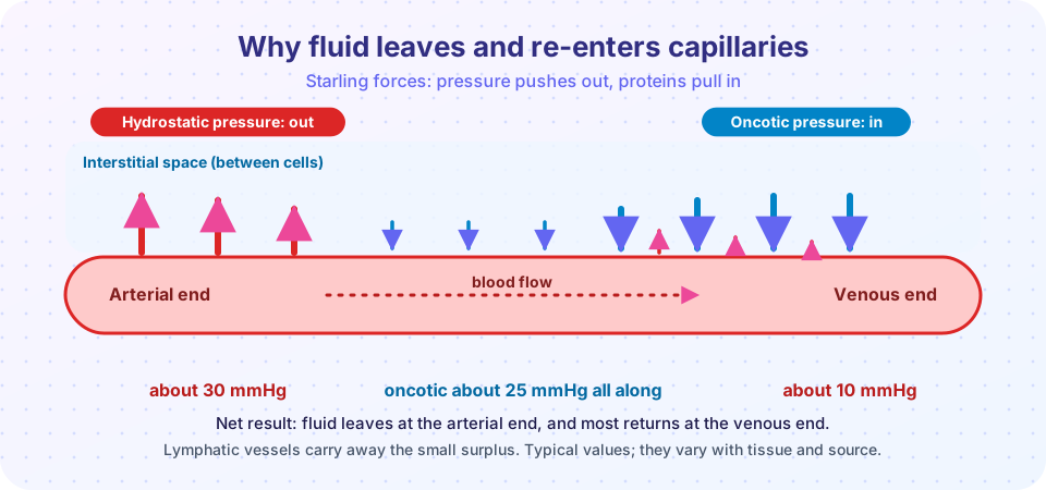
Water moves across the capillary wall in response to four pressures:
- Capillary hydrostatic pressure pushes fluid out. It is higher at the arterial end than the venous end.
- Plasma oncotic (colloid osmotic) pressure pulls fluid in. It is created mainly by the protein albumin, which is too large to leave the vessel.
- Interstitial hydrostatic pressure, which is small.
- Interstitial oncotic pressure, which is small because tissue fluid has little protein.
The net movement is approximately (capillary hydrostatic - interstitial hydrostatic) - (plasma oncotic - interstitial oncotic). In a healthy capillary, outward filtration at the arterial end is balanced by inward reabsorption at the venous end, and the lymphatic vessels carry the small leftover back to the blood. The exact numbers vary from tissue to tissue.
Edema appears when one or more of these forces is altered.
The five causes
| Cause | Mechanism | Example |
|---|---|---|
| Increased hydrostatic pressure | Venous pressure backs up and pushes more fluid out | Heart failure, a deep vein blockage, liver cirrhosis (portal hypertension causes ascites) |
| Reduced plasma oncotic pressure | Too little albumin to hold water in | Nephrotic syndrome (protein lost in urine), liver disease (low synthesis), severe malnutrition |
| Lymphatic obstruction | Fluid cannot drain away | Surgical removal of lymph nodes, radiation, tumor, filarial parasites |
| Sodium and water retention | Extra volume raises hydrostatic pressure and dilutes protein | Kidney disease, excess aldosterone, high salt intake in susceptible people |
| Increased vascular permeability | Leaky vessels let protein out (exudate) | Inflammation, burns |
Several often work together. In heart failure, the failing heart raises venous pressure and the kidneys retain sodium and water in response to reduced flow, so more than one mechanism is at work.
What edema looks like
Edema is described by where it is. In subcutaneous tissue it is often gravity-dependent: ankle swelling in a person who is standing all day, or sacral swelling in someone confined to bed. Pressing a finger into the swollen area leaves a dent, called pitting edema. In pulmonary edema the lungs are heavy and wet, and frothy fluid fills the air spaces, causing breathlessness. In brain edema, the brain swells within the rigid skull; the folds flatten, and increased pressure can push the brain through openings in the skull (herniation), which is life-threatening.
Fluid in a body cavity is called an effusion. Severe, generalized edema is called anasarca.
Hyperemia and congestion
Both mean "more blood than usual in a tissue," but the cause and the color differ.
- Hyperemia is active: arterioles widen and more blood flows in. The tissue is red. Examples are blushing, exercising muscle and acute inflammation.
- Congestion is passive: venous outflow is blocked or slowed, so blood pools. The tissue is blue-red (cyanotic) because the trapped blood has lost its oxygen. Causes include heart failure and local obstruction.
Chronic congestion is not harmless. In chronic passive congestion of the lung caused by left-sided heart failure, the lungs are stiff and brownish because red cells leak into the air spaces and are taken up by macrophages ("heart failure cells", full of iron-containing hemosiderin). In congestion of the liver from right-sided heart failure, the center of each lobule suffers most from low oxygen, so the cut surface shows dark red centers and paler edges, the "nutmeg liver".
Hemorrhage
Hemorrhage is the escape of blood from a vessel. It may be external, or hidden inside the body.
| Term | Meaning |
|---|---|
| Hematoma | A collection of blood within a tissue (a bruise is a small one) |
| Petechiae | Pinpoint hemorrhages, 1 to 2 mm, in skin or mucous membranes |
| Purpura | Spots larger than 3 mm |
| Ecchymosis | Larger bruise, more than 1 to 2 cm |
| Hemothorax, hemopericardium, hemoperitoneum, hemarthrosis | Blood in the chest cavity, heart sac, abdomen and a joint |
A bruise changes color because the hemoglobin from the escaped red cells is broken down in stages: red-blue, then green (biliverdin), then yellow (bilirubin), then golden brown (hemosiderin).
How serious a bleed is depends on volume, rate and site. A slow loss of a large volume may be tolerated; rapid loss of 20 percent or more of blood volume can cause shock. Even a small amount may be fatal in a critical place, such as bleeding into the brain or the sac around the heart. Chronic external blood loss, for instance from an ulcer, causes iron deficiency anemia.
Hemostasis: how bleeding stops
When a vessel is cut, two connected stages work together.
Primary hemostasis builds the platelet plug within seconds to minutes:
- The vessel constricts reflexively.
- Platelets stick to exposed collagen through a bridge protein, von Willebrand factor.
- Platelets are activated, change shape, release granule contents and recruit more platelets, which clump together (aggregation) to form the plug.
Secondary hemostasis is the coagulation cascade. The exposed tissue factor from the damaged wall triggers a sequence of clotting factors that ends with thrombin, which converts soluble fibrinogen into fibrin threads. Fibrin makes the platelet plug firm and permanent. Many clotting factors need vitamin K to be made correctly. Normal blood also has built-in brakes: antithrombin, protein C and protein S limit clotting, and the enzyme plasmin dissolves clots after healing (fibrinolysis). Balance is the goal: too little clotting means bleeding; too much means thrombosis (the next lesson).
Bleeding disorders
- Vessel wall defects: scurvy (vitamin C needed for strong collagen), inflammation of vessels (vasculitis), some infections.
- Platelet disorders: low platelet count (thrombocytopenia) or poorly functioning platelets, often leading to petechiae and mucosal bleeding.
- Clotting factor deficiencies: hemophilia A (factor VIII), hemophilia B (factor IX), and von Willebrand disease. These cause deep bleeding into joints and muscles. Hemophilia A and B are X-linked and thus mainly affect males.
- Acquired causes: liver disease (fewer clotting factors made), vitamin K deficiency and anticoagulant drugs such as warfarin or heparin.
Common misconceptions
- "Edema is always caused by too much salt." Salt intake can contribute, but heart, kidney, liver and lymphatic diseases are major causes. Swelling needs medical evaluation.
- "Pitting edema means a vein problem only." It reflects fluid retention from any of several mechanisms.
- "A bruise is a bleeding disorder." Easy bruising can result from minor bumps, age or medications; only a pattern of unusual bleeding points to a disorder.
Try it
The first block calculates the net filtration at the two ends of a capillary under four conditions. The values are simplified textbook numbers used to show the logic, not measurements.
Press Run (or Ctrl + Enter) to see the result here.
Program input · what you'd type while the program runs
Type the answers before you press Run, one per line. Each time the program reads a line (with input()), it gets the next line from this box.
The second block names a hemorrhage by the size of its spots. The size cut-offs are the usual textbook ones.
Press Run (or Ctrl + Enter) to see the console output here.
No output.
Check your understanding
Quick check · 6 questions
of 6 answered You scored / 6 · perfect!
-
1. Which plasma protein mainly provides oncotic pressure?
Albumin is the most abundant plasma protein and holds water in the vessels. -
2. Which condition causes edema mainly by reducing plasma oncotic pressure?
In nephrotic syndrome, albumin is lost in the urine. -
3. What distinguishes congestion from hyperemia?
Congestion is passive and bluish; hyperemia is active and red. -
4. Pinpoint hemorrhages 1 to 2 mm across are called:
Petechiae are the smallest; purpura are larger than 3 mm; ecchymoses larger than 1 cm. -
5. Which protein bridges platelets to exposed collagen?
von Willebrand factor links platelet receptors to collagen in primary hemostasis. -
6. Hemophilia A is caused by deficiency of which factor?
Factor VIII deficiency causes hemophilia A, an X-linked disorder.
Summary
- Fluid exchange depends on hydrostatic pressure (out) and oncotic pressure (in), with lymphatics draining the surplus.
- Edema has five main causes: raised venous pressure, low albumin, lymphatic obstruction, sodium and water retention, and increased permeability.
- Hyperemia is active and red; congestion is passive and bluish, with nutmeg liver and heart failure cells as classic examples.
- Hemorrhage is named by size and site; its seriousness depends on volume, rate and location.
- Hemostasis has a platelet stage and a coagulation stage; deficiencies in vessels, platelets or clotting factors cause bleeding.
Track your progress
Create a free account to tick off lessons and pick up where you left off on any device.
of Pathology lessons completed