


















Lesson 29 of 46 · Cardiovascular and Respiratory Pathology
Lung disease: obstructive, restrictive, pneumonia and cancer
Compare obstructive and restrictive lung disease with spirometry, then study emphysema, asthma, pulmonary fibrosis, pneumonia patterns and the main types of lung cancer.
Every breath is a negotiation between two physical properties: how easily air flows through the airways, and how easily the lungs stretch. Disease can disturb either one. If air cannot flow out freely, the disease is obstructive. If the lungs cannot expand, it is restrictive. This simple split, made visible by a breathing test called spirometry, organizes most of non-infectious lung pathology.
This lesson starts with that split, goes through the key obstructive and restrictive conditions, and then turns to two of the most important lung problems of all: infection and cancer.
What you will learn
- How spirometry separates obstructive from restrictive patterns
- The pathology of emphysema, chronic bronchitis and asthma
- Interstitial (restrictive) disease, such as fibrosis and pneumoconiosis
- The patterns of pneumonia
- The main types of lung cancer and their links to smoking
Key terms
FEV1: forced expiratory volume in the first second of a hard exhalation.
FVC: forced vital capacity, the total volume blown out.
Emphysema: permanent enlargement of air spaces beyond the terminal bronchioles with destruction of their walls.
Fibrosis: scarring with excess collagen.
Consolidation: lung tissue filled with fluid or cells, so that it is solid instead of air-filled.
Spirometry in one picture
In a spirometry test a person breathes in fully and blows out as hard and as long as possible. The machine records volume against time. Two numbers matter most: the FVC, and the FEV1 (what was blown out in the first second). The FEV1/FVC ratio shows how fast the air comes out.
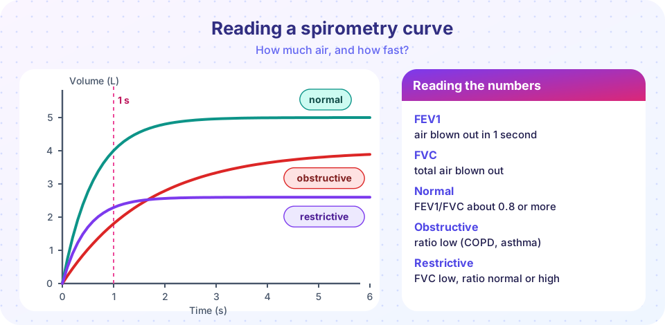
- In obstructive disease air is slow to leave, so FEV1 falls much more than FVC and the ratio is low. A commonly used fixed threshold is a ratio below 0.7 after a bronchodilator.
- In restrictive disease the lungs are stiff or small, so both FEV1 and FVC fall but air still leaves quickly: FVC is low and the ratio is normal or even high.
A caution: spirometry suggests a pattern, and diagnosing a restrictive disease usually needs lung volume measurements and imaging as well.
Press Run (or Ctrl + Enter) to see the result here.
Program input · what you'd type while the program runs
Type the answers before you press Run, one per line. Each time the program reads a line (with input()), it gets the next line from this box.
Obstructive lung disease
Emphysema
Emphysema is the permanent loss of the walls between air sacs, so many small alveoli merge into a few large ones. Surface for gas exchange shrinks, and the lung loses its elastic recoil, so during exhalation small airways collapse and trap air. Patients are breathless, with an over-inflated "barrel" chest.
The mechanism is an imbalance between enzymes that digest elastin (released by neutrophils and macrophages) and protective antiprotease. Smoke recruits inflammatory cells and also inactivates the protective alpha-1 antitrypsin. Two patterns show the logic:
- Centriacinar emphysema begins around the respiratory bronchioles, mainly in the upper lobes, and is the typical pattern in cigarette smokers.
- Panacinar emphysema involves the whole acinus, mainly in the lower lobes, and is characteristic of inherited alpha-1 antitrypsin deficiency, which can occur even in non-smokers.
Press Run (or Ctrl + Enter) to see the result here.
Program input · what you'd type while the program runs
Type the answers before you press Run, one per line. Each time the program reads a line (with input()), it gets the next line from this box.
Chronic bronchitis
Chronic bronchitis is defined clinically as a productive cough on most days for at least three months in two consecutive years. The airways show enlarged mucus-secreting glands, mucus plugging, and inflammation. It is mostly caused by smoke. Emphysema and chronic bronchitis usually coexist, and together they are called chronic obstructive pulmonary disease (COPD).
Asthma
Asthma is a chronic inflammatory disease of the airways with reversible narrowing. In the common allergic form, an inhaled allergen triggers a Th2-type immune response, with IgE, mast cells and eosinophils (a type I hypersensitivity). The airways become over-reactive: smooth muscle contracts, mucus is secreted, and the wall swells, which causes wheezing, cough and breathlessness. Pathology shows thick mucus plugs, thickened basement membrane, enlarged smooth muscle and many eosinophils. Dense mucus can contain Curschmann spirals (coiled mucus casts) and Charcot-Leyden crystals (from eosinophil proteins). Over many years, the airway can remodel, and part of the narrowing becomes fixed.
Bronchiectasis, the permanent dilation of bronchi after chronic infection or blockage, is a third obstructive disease, classically seen in cystic fibrosis.
Restrictive lung disease
Restriction has two main kinds of cause: chest wall problems (obesity, scoliosis, neuromuscular disease) and interstitial lung disease, which scars or inflames the delicate tissue between the air sacs.
- Idiopathic pulmonary fibrosis (IPF) is progressive scarring with no known cause, a pattern called usual interstitial pneumonia. Pathology shows patchy fibrosis, fibroblast foci and honeycomb cysts, with a preference for the lung bases and the periphery. Patients develop a dry cough and progressive breathlessness. Scarring probably results from repeated epithelial injury followed by exaggerated repair.
- Pneumoconioses are lung diseases from inhaled dust. Silicosis (from silica in mining, stone cutting and sandblasting) and coal workers' pneumoconiosis affect mainly the upper lobes. Asbestosis causes basal fibrosis with ferruginous bodies (asbestos fibers coated with iron), plaques on the pleura, and a risk of lung cancer and mesothelioma.
- Sarcoidosis is a multisystem disease with non-caseating granulomas, often in the lungs and lymph nodes in the chest.
- Hypersensitivity pneumonitis is an immune reaction to inhaled organic dust, such as moldy hay.
- Acute respiratory distress syndrome (ARDS) is acute, widespread injury of the alveolar walls, from sepsis, trauma, aspiration or infection, with leaky capillaries and hyaline membranes lining the air spaces.
Pneumonia
Pneumonia is infection of the lung's air spaces. The pattern tells you about the organism and the route.
- Lobar pneumonia fills an entire lobe, classically from Streptococcus pneumoniae. Four stages are described: congestion (vascular engorgement, fluid), red hepatization (red cells and neutrophils fill the air spaces so the lung feels like liver), gray hepatization (red cells break down; the lung is gray-brown with fibrin and neutrophils), and resolution (enzymes digest the exudate). Antibiotics change the course, so the full sequence is rarely seen now.
- Bronchopneumonia is patchy, centered on airways, and common in the very young, old and ill, caused by organisms such as Staphylococcus aureus, Haemophilus and Pseudomonas.
- Interstitial ("atypical") pneumonia involves the walls of the air sacs, caused by Mycoplasma, viruses such as influenza, and others.
- Aspiration pneumonia follows inhaled stomach contents or mouth bacteria, typically in the lower, right-sided lung segments, and can form abscesses.
Complications include lung abscess, empyema (pus in the pleural space) and spread to the blood. The causes differ in hospitalized patients, where resistant bacteria are more likely.
Lung cancer
Lung cancer is the leading cause of cancer death in much of the world. Cigarette smoking accounts for the large majority of cases, and the risk depends on how much and for how long a person has smoked. Other causes are radon gas, asbestos, other occupational exposures and air pollution. Lung cancers arise by multistep carcinogenesis, as in the previous chapter, with mutations in genes such as TP53, KRAS and EGFR.
Cancers are divided mainly into small cell and non-small cell types, because their treatment differs.
| Type | Typical features |
|---|---|
| Adenocarcinoma | Most common overall now; usually peripheral; also the main type in non-smokers; often has driver mutations (EGFR, ALK) that can be targeted by drugs |
| Squamous cell carcinoma | Often central, near the large bronchi; strongly linked to smoking; keratin and cavitation; may secrete PTHrP, causing high calcium |
| Small cell carcinoma | Central; neuroendocrine cells with scant cytoplasm; very aggressive; early spread; may secrete ACTH or ADH |
| Large cell carcinoma | Poorly differentiated, a diagnosis of exclusion |
A tumor at the lung apex (a Pancoast tumor) can invade nerves and cause shoulder pain and Horner syndrome. Screening with low-dose CT is recommended in some countries for heavy smokers, because early-stage disease is much more treatable.
Common misconceptions
- "Only smokers get emphysema or lung cancer." Smoking is the main cause, but alpha-1 antitrypsin deficiency, radon, occupational dusts, air pollution and genetics also contribute; adenocarcinoma occurs in non-smokers.
- "Asthma and COPD are the same." Asthma is typically reversible and allergic; COPD is largely irreversible and smoke related, though they can overlap.
- "Pneumonia is always bacterial." Viruses and other organisms are also common causes.
Try it
Press Run (or Ctrl + Enter) to see the console output here.
No output.
Check your understanding
Quick check · 6 questions
of 6 answered You scored / 6 · perfect!
-
1. Which spirometry result suggests an obstructive pattern?
In obstruction air leaves slowly, so FEV1 falls out of proportion to FVC. -
2. Which type of emphysema is typical of alpha-1 antitrypsin deficiency?
Panacinar emphysema with a lower lobe predominance is characteristic. -
3. Charcot-Leyden crystals and Curschmann spirals are linked to
They are found in the mucus of asthma, which is rich in eosinophils. -
4. The red hepatization stage of lobar pneumonia is characterized by
In red hepatization the air spaces are packed with red cells, neutrophils and fibrin, and the lung feels solid. -
5. Which lung cancer is typically central, aggressive, and may secrete ACTH?
Small cell carcinoma is a neuroendocrine tumor that spreads early and can make hormones. -
6. Honeycomb fibrosis at the lung bases is typical of
IPF (usual interstitial pneumonia) causes subpleural basal fibrosis with honeycomb cysts.
Summary
- Obstructive disease slows exhalation (low FEV1/FVC); restrictive disease limits expansion (low FVC, normal or high ratio).
- Emphysema destroys alveolar walls; chronic bronchitis is a clinical syndrome of mucus hypersecretion; asthma is reversible allergic airway inflammation.
- Interstitial fibrosis and pneumoconioses are the main restrictive diseases.
- Pneumonia can be lobar, bronchopneumonia, interstitial or aspiration.
- Lung cancer is divided into small cell and non-small cell, with smoking the dominant cause.
Next: Chapter review
Track your progress
Create a free account to tick off lessons and pick up where you left off on any device.
of Pathology lessons completed