


















Lesson 8 of 46 · Cell Injury and Death
Reversible and Irreversible Cell Injury: From Low Oxygen to Cell Death
Follow low oxygen through ATP loss, swelling, calcium overload and free radicals to the point of no return, and learn how reperfusion and toxins injure cells.
Imagine switching off the oxygen supply to a heart muscle cell. For a short time nothing seems to happen. Then the cell swells, its energy stores drop and its membranes begin to leak. If the oxygen returns quickly enough, the cell recovers; if not, it crosses a boundary after which no treatment can save it. This lesson follows that sequence step by step. It is the model for most injury in pathology: understand it once with oxygen, and you can see the same logic behind poisons, infections and many diseases of organs.
What you will learn
- The chain of events from oxygen loss to swelling and, in the end, to death.
- What the cell looks like in reversible injury, and what marks irreversibility.
- How free radicals, calcium and misfolded proteins contribute.
- Why restoring blood flow can sometimes cause extra damage.
- How leaked cell contents become blood tests.
Key terms
ATP: adenosine triphosphate, the cell's energy currency.
Hydropic change (cellular swelling): the earliest sign of many injuries, caused by water entering the cell.
Reactive oxygen species (ROS): chemically aggressive oxygen-containing molecules, such as superoxide and hydrogen peroxide, that damage lipids, proteins and DNA.
Reperfusion injury: extra damage that can follow restoration of blood supply to ischemic tissue.
Mitochondrial permeability transition: opening of a large pore in the mitochondrial membrane that collapses energy production.
Step 1: the energy crisis
Most ATP in a healthy cell is made by the mitochondria through oxidative phosphorylation, which needs oxygen. When oxygen runs low, ATP production falls. The cell shifts to anaerobic glycolysis, which makes some ATP without oxygen but is far less efficient and produces lactic acid. Falling ATP has several immediate consequences:
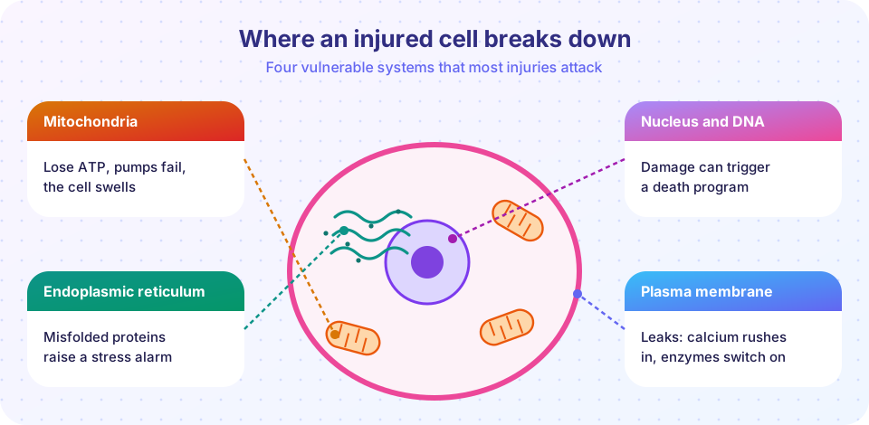
- The sodium pump fails. The Na⁺/K⁺-ATPase uses ATP to push sodium out of the cell and potassium in. Without ATP, sodium and water accumulate inside. The cell swells and the endoplasmic reticulum dilates. This is the first visible change: cellular swelling.
- Glycogen is used up and lactic acid builds. The intracellular pH drops, causing clumping of the nuclear chromatin.
- Ribosomes detach from the rough endoplasmic reticulum and protein synthesis falls.
- Membrane function declines, with blebs forming on the surface.
At this stage a cell has changed but is still intact. The damage is potentially reversible: if oxygen is restored, the pump restarts, water leaves and the cell returns to normal.
What reversible injury looks like
Two patterns are especially important.
- Cellular swelling. The cell is larger and paler. In a whole organ the result is pallor, increased weight and a tense surface. The change is subtle at low power: the finding that cells are bigger with a clear or granular cytoplasm may be all that a pathologist sees.
- Fatty change (steatosis). In cells that handle a lot of fat, especially liver cells, injury from toxins, alcohol, diabetes or obesity can lead to small or large lipid droplets accumulating in the cytoplasm. It is common and often reversible.
Other features seen under the electron microscope include blebs of the plasma membrane, swollen mitochondria and the formation of whorled structures called myelin figures, derived from damaged membranes.
The point of no return
What turns reversible into irreversible injury? Cell biologists agree on two features that are consistently associated with death:
- Inability to reverse mitochondrial dysfunction. Even when oxygen is restored, the mitochondria cannot restart ATP production. A key event is the opening of a large pore in the inner mitochondrial membrane (the permeability transition), which dissipates the proton gradient that drives ATP synthesis.
- Profound disturbances in membrane function. The plasma membrane and lysosome membranes become leaky.
Calcium plays an important role. In a healthy cell the free calcium inside is kept extremely low, about ten thousand times lower than outside. When ATP fails, pumps that remove calcium cannot work and calcium flows in from outside and from internal stores. High calcium activates a collection of enzymes: phospholipases (which damage membranes), proteases (which digest the cytoskeleton), endonucleases (which cut DNA) and ATPases (which consume the remaining ATP). It also promotes the mitochondrial permeability transition. Lysosomes then leak digestive enzymes into the cytoplasm, and the cell is digested from within.
The time needed to cross the boundary depends on the cell. In human brain tissue permanent injury begins within a few minutes of complete circulatory arrest. In heart muscle, experiments suggest irreversible damage in the center of a blocked artery's territory starts after roughly twenty to forty minutes of severe ischemia. Skeletal muscle and the kidney tolerate longer. These time windows explain why rapid reopening of a blocked coronary artery saves heart muscle.
Free radicals and oxidative stress
Oxygen is life-giving, but its partly reduced forms are dangerous. Reactive oxygen species include superoxide (O₂⁻), hydrogen peroxide (H₂O₂) and the very reactive hydroxyl radical (•OH). They are produced normally in small amounts, for instance as a byproduct of mitochondrial respiration, and are used by white cells to kill microbes. They are also produced in excess by radiation, certain chemicals, inflammation and the return of oxygen after ischemia.
ROS injure cells in three main ways: lipid peroxidation of membranes, oxidation of proteins (which damages enzymes and promotes misfolding) and DNA damage (breaks and base changes). Cells defend themselves with antioxidants and enzymes: superoxide dismutase turns superoxide into hydrogen peroxide; catalase and glutathione peroxidase convert hydrogen peroxide into water. Vitamins E and C and glutathione scavenge radicals. Injury occurs when production outpaces defense, a state called oxidative stress.
Reperfusion injury
It seems obvious that restoring blood flow can only help. For cells that are already irreversibly injured, nothing changes. But for cells that are on the border, reperfusion can itself add damage. Returning oxygen generates a burst of ROS because the damaged mitochondria and enzymes handle it poorly. Calcium overload may worsen. Inflammatory cells and complement proteins arrive with the blood. The result can be extra cell death beyond that of the original ischemia. This phenomenon is called ischemia-reperfusion injury. It is a reason why research seeks drugs that can limit it, and why timing of intervention matters. It does not mean reperfusion should be avoided: in heart attack and stroke, early restoration of flow remains the most effective way to save tissue.
Other injury paths
- Misfolded proteins and ER stress. The endoplasmic reticulum folds and checks new proteins. If misfolded proteins accumulate (from genetic mutations, aging, oxidative damage or a viral infection), the cell activates the unfolded protein response to slow synthesis and improve folding. If the stress is overwhelming, the response switches to promoting apoptosis. This mechanism is involved in diseases like cystic fibrosis and some neurodegenerative diseases.
- Chemical injury. Some chemicals act directly, such as mercury binding to enzymes. Many are harmless until the liver converts them: carbon tetrachloride and an overdose of acetaminophen (paracetamol) are changed by cytochrome P450 enzymes into toxic products that attack membranes and proteins.
- Infectious and immune injury will be discussed in the chapters on inflammation and infection.
Dead cells leak: the basis of blood tests
When the plasma membrane breaks, intracellular proteins leak into the blood. Their levels act as markers of injury in specific organs. Troponin (a heart muscle protein) rises after heart muscle injury. The liver enzymes ALT and AST rise in liver cell injury. These tests exist because cell death is not silent: contents spill, and the blood reports on it.
Common misconceptions
- "Cells die immediately when oxygen stops." Cells tolerate hypoxia for minutes to hours, depending on type, and early injury is reversible.
- "Reperfusion is dangerous and should be avoided." Early restoration of flow is the best treatment; reperfusion injury is a limiting effect that scientists try to reduce.
- "Swelling means the cell is dead." Swelling is the earliest sign of reversible injury.
Try it
The first block is a toy simulation. ATP falls during ischemia, the cell swells because the sodium pump fails, and a threshold mimics the point of no return. The parameters are invented for teaching, not measured values.
Press Run (or Ctrl + Enter) to see the result here.
Program input · what you'd type while the program runs
Type the answers before you press Run, one per line. Each time the program reads a line (with input()), it gets the next line from this box.
The second block is a lookup of the blood markers used to detect injury in different organs. It lists the usual textbook pairings; it is for learning, not for interpreting any real result.
Press Run (or Ctrl + Enter) to see the console output here.
No output.
Check your understanding
Quick check · 6 questions
of 6 answered You scored / 6 · perfect!
-
1. What is the first visible change in many reversible injuries?
When the sodium pump fails, water enters and the cell swells. -
2. Why does the sodium pump fail during hypoxia?
The Na+/K+ ATPase requires ATP, which oxidative phosphorylation can no longer supply. -
3. Which two features are consistently linked to irreversible injury?
Failure to restore ATP production and profound membrane dysfunction mark the point of no return. -
4. How do high intracellular calcium levels cause damage?
Calcium activates destructive enzymes and promotes the mitochondrial permeability transition. -
5. Which enzyme converts hydrogen peroxide into water?
Catalase and glutathione peroxidase convert hydrogen peroxide into water; superoxide dismutase makes hydrogen peroxide from superoxide. -
6. A patient's blood troponin rises after chest pain. Which is the most likely explanation?
Troponin is a heart muscle protein; its appearance in blood signals membrane damage in heart cells.
Summary
- Low oxygen lowers ATP, so the sodium pump fails, water enters, and the cell swells: reversible injury.
- Fatty change, membrane blebs and swollen mitochondria are other reversible signs.
- Irreversibility is marked by mitochondrial failure that cannot recover and by severe membrane damage, with calcium overload and enzyme activation.
- Reactive oxygen species damage lipids, proteins and DNA; antioxidant enzymes and vitamins protect.
- Reperfusion can add injury to borderline cells, but early reperfusion is still the best therapy.
- Leaked cell proteins make injury measurable in blood tests.
Next: Necrosis and Apoptosis
Track your progress
Create a free account to tick off lessons and pick up where you left off on any device.
of Pathology lessons completed