


















Lesson 10 of 46 · Cell Injury and Death
Chapter 2 Review: Cell Injury and Death
Review cell injury: causes, adaptations, reversible versus irreversible injury, necrosis patterns and apoptosis, with a recap table and a ten-question quiz.
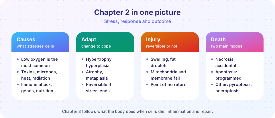
Chapter 2 gave you the central story of cell pathology. A stress arrives; the cell adapts, is injured but recovers, or dies; and the manner of death shapes what the tissue does next. Everything in the chapters ahead, from inflammation to tumors, builds on this storyline. Use this review to test yourself, not just to reread. Try to explain each row of the recap table aloud before looking at it.
What you will learn
- How the four lessons of chapter 2 connect.
- The terms and mechanisms that you should be able to recall.
- Which parts of the chapter need another pass, through a mixed quiz.
Recap table
| Topic | Key facts |
|---|---|
| Causes | Hypoxia and ischemia, physical, chemical, infectious, immune, genetic, nutritional, aging |
| Vulnerable systems | ATP and mitochondria, membranes, protein folding (ER), DNA |
| Adaptations | Hypertrophy (size up), hyperplasia (number up), atrophy (size down), metaplasia (type changed) |
| Reversible injury | ATP falls, sodium pump fails, swelling, fatty change, blebs |
| Irreversible injury | Mitochondrial failure that cannot recover, membrane damage, calcium overload, enzyme release |
| Free radicals | Superoxide, hydrogen peroxide, hydroxyl radical; defenses: SOD, catalase, glutathione peroxidase, vitamins E and C |
| Necrosis | Membrane rupture and inflammation; pyknosis, karyorrhexis, karyolysis; coagulative, liquefactive, caseous, fat, fibrinoid |
| Apoptosis | Programmed, energy-dependent, no inflammation; BCL-2 family, cytochrome c, caspases 3, 6, 7; death receptors and caspase-8 |
Three questions to ask of any slide or case
- Is the tissue adapting, injured or dead? (Size, number, type, nuclei.)
- If injured or dead, which system failed first? (Energy, membrane, proteins, DNA.)
- If dead, was it necrosis or apoptosis, and which pattern? What does that say about the cause?
Worked mini-cases
Case 1: the long flight and the cast. A leg is held in a plaster cast for six weeks. The muscle is smaller afterwards. Which adaptation? Atrophy: the cells shrink through decreased protein synthesis and increased breakdown by the proteasome and autophagy. The cells are alive, and with use the muscle will hypertrophy back.
Case 2: the blocked artery. Blood flow to part of the heart stops. In the first minutes ATP falls, the sodium pump fails and the cells swell (reversible). If flow is restored early, the cells recover. If the block lasts beyond a critical time, mitochondria cannot restart, membranes leak, calcium floods in and cell death follows. Troponin appears in the blood. The pattern in the tissue is coagulative necrosis, and inflammation follows. Notice how every idea from the chapter appears in a single story.
Case 3: the shrinking nucleus. On a slide, a cell has a dark, shrunken nucleus and the cell is in a clear space with no inflammation around it. Which kind of death? The lack of inflammation and the shrunken cell suggest apoptosis; a pyknotic nucleus with a swollen, pink cell and inflammation around it would suggest necrosis. Always combine cell features with the surrounding tissue.
Try it
The first block is a flashcard drill for the vocabulary of the chapter.
Press Run (or Ctrl + Enter) to see the console output here.
No output.
The second block draws the comparison table for necrosis and apoptosis as a figure you can adapt as a revision aid.
Press Run (or Ctrl + Enter) to see the result here.
Program input · what you'd type while the program runs
Type the answers before you press Run, one per line. Each time the program reads a line (with input()), it gets the next line from this box.
Check your understanding
Quick check · 10 questions
of 10 answered You scored / 10 · perfect!
-
1. Which is the most common cause of cell injury?
Hypoxia and ischemia are the most frequent causes of cell injury in disease. -
2. A weightlifter's biceps grows larger because the muscle cells enlarge. This is:
Larger cells with the same number is hypertrophy. -
3. Barrett esophagus is an example of:
The normal squamous lining is replaced by glandular columnar cells in response to chronic acid reflux. -
4. Which event directly causes cellular swelling in early ischemia?
Without ATP the pump cannot remove sodium, so water follows into the cell. -
5. Which is NOT a feature of irreversible injury?
Swelling alone is reversible; irreversibility involves mitochondria and membranes. -
6. Which defence enzyme turns superoxide into hydrogen peroxide?
Superoxide dismutase does this; catalase and glutathione peroxidase then remove hydrogen peroxide. -
7. Which pattern of necrosis is typical of a bacterial abscess?
Enzymes from white cells dissolve tissue into pus. -
8. Which is true of apoptosis?
Apoptosis is an energy-dependent program carried out by caspases and is essential in development. -
9. Which molecule leaks from mitochondria to start the intrinsic apoptosis pathway?
Cytochrome c joins APAF-1 to form the apoptosome and activate caspase-9. -
10. Too little apoptosis is associated with:
When abnormal or self-reactive cells fail to die they can accumulate.
Summary
- Cells adapt, recover from reversible injury, or die, depending on stress, cell type and history.
- Hypertrophy, hyperplasia, atrophy and metaplasia are the four adaptations.
- Energy loss, sodium pump failure and swelling begin reversible injury; mitochondrial and membrane failure marks the point of no return.
- Necrosis is messy and inflammatory with five patterns; apoptosis is programmed and silent.
Track your progress
Create a free account to tick off lessons and pick up where you left off on any device.
of Pathology lessons completed