


















Lesson 38 of 46 · Infectious and Neurological Disease
Stroke and dementia: cerebrovascular and neurodegenerative disease
Understand ischemic and hemorrhagic stroke, what happens to the brain after an infarct, and the protein pathology of Alzheimer, Lewy body, Parkinson and Huntington disease.
The brain is about two percent of body weight but uses roughly a fifth of the body's oxygen. It has almost no reserve, so when blood supply is cut off, brain tissue begins to die within minutes. That makes stroke one of the great causes of death and disability. Over a longer timescale, a different problem threatens the brain: neurons may be lost slowly as misfolded proteins accumulate, producing dementia and movement disorders.
The two topics are linked, because small strokes are a leading cause of dementia and because vascular disease and Alzheimer pathology often appear together in older brains. This lesson takes each in turn.
What you will learn
- The difference between ischemic and hemorrhagic stroke and their causes
- How an infarct in the brain evolves (and why it differs from one in the heart)
- The protein lesions of Alzheimer disease and how they spread
- How Lewy body disease, Parkinson disease, frontotemporal dementia, Huntington disease and prion disease differ
Key terms
Infarct / penumbra: dead tissue / surrounding tissue that is at risk but may be saved.
Liquefactive necrosis: tissue death in which dead cells dissolve into a liquid, typical of the brain.
Gliosis: proliferation of astrocytes, the brain's form of scar.
Amyloid / tau: proteins that form plaques outside / tangles inside neurons in Alzheimer disease.
Prion: a misfolded protein that spreads by converting normal copies.
Stroke
A stroke is a sudden neurological deficit caused by a vascular problem. Doctors use a quick public mnemonic, "FAST" (face drooping, arm weakness, speech difficulty, time to call emergency services), because the faster blood flow is restored the more brain survives.
Ischemic stroke
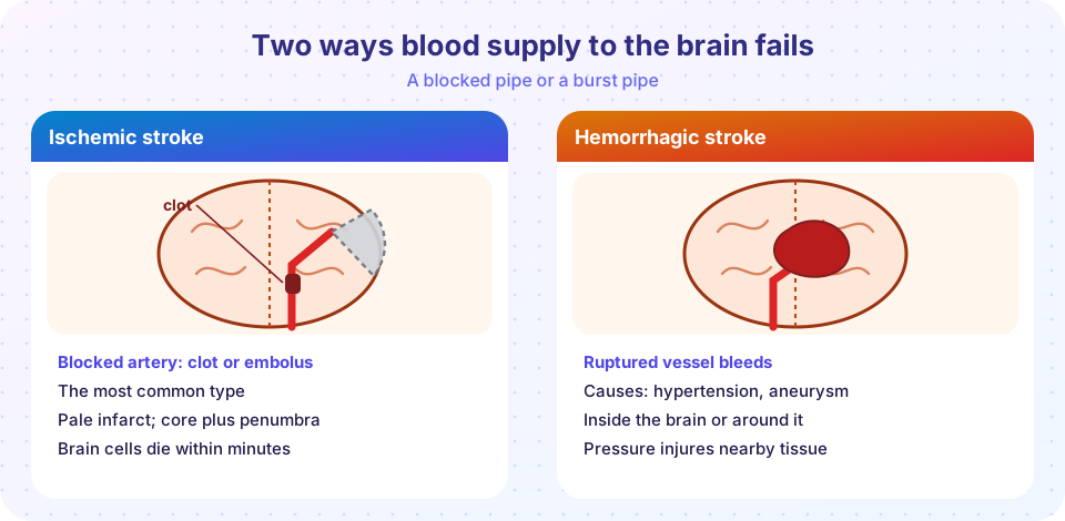
Most strokes are ischemic, caused by blockage of an artery. The blockage may be:
- Thrombotic: a clot forms on an atherosclerotic plaque in a brain artery, such as the middle cerebral artery.
- Embolic: a clot or fragment forms elsewhere and travels to the brain. The heart is the most important source, especially in atrial fibrillation, where blood pools in the atrium and clots, as are plaques in the carotid artery of the neck.
- Small vessel (lacunar): hypertension damages the small deep arteries, causing tiny infarcts (lacunes), which in large numbers cause cognitive decline.
- Global hypoperfusion: a drop in blood pressure (cardiac arrest, shock) damages the border zones between major arteries (watershed infarcts).
A transient ischemic attack (TIA) is a brief episode of the same symptoms without permanent infarction, and it is a warning sign that a full stroke may follow.
What happens to the tissue
In the core of the infarct, cells die within minutes. Around it lies the penumbra, tissue with reduced flow that is functionally silent but still alive, and which can be saved if flow returns soon. This is the basis of the phrase "time is brain": a widely quoted estimate is that a large-vessel stroke destroys on the order of two million neurons every minute that it is untreated.
The brain infarct changes with time in a pattern unlike the heart's coagulative necrosis. Brain tissue is rich in lipid and poor in structural protein, so dead tissue becomes liquefactive necrosis.
- 12 to 24 hours: shrunken, red (eosinophilic) neurons.
- 24 to 72 hours: neutrophils.
- Days to weeks: macrophages (microglia) full of lipid, called foamy cells, remove dead tissue, and capillaries grow.
- Weeks to months: a fluid-filled cavity surrounded by gliosis by astrocytes. The brain does not form a collagen scar the way other organs do.
Press Run (or Ctrl + Enter) to see the result here.
Program input · what you'd type while the program runs
Type the answers before you press Run, one per line. Each time the program reads a line (with input()), it gets the next line from this box.
Hemorrhagic stroke and other bleeding
- Intracerebral hemorrhage is bleeding into brain tissue. The commonest cause is hypertension, which damages small penetrating arteries in the deep structures (basal ganglia, thalamus, pons, cerebellum). In older adults cerebral amyloid angiopathy, the deposition of amyloid in vessel walls, is a cause of bleeding in the outer cortex (lobar hemorrhage).
- Subarachnoid hemorrhage is bleeding into the space around the brain, usually from a ruptured berry aneurysm at the circle of Willis (a bulge at an arterial branch point). The classic symptom is a sudden, severe headache, often described as the worst of the person's life. Complications are rebleeding, vasospasm and hydrocephalus. Polycystic kidney disease and connective tissue disorders increase aneurysm risk.
- Epidural hematoma follows trauma with a skull fracture that tears the middle meningeal artery. Blood collects between the skull and the dura, forming a lens-shaped mass, often with a "lucid interval" before deterioration.
- Subdural hematoma comes from torn bridging veins, producing a crescent-shaped collection, common in older adults and alcohol-dependent people after minor head injury.
Dementia
Dementia is an acquired, usually progressive decline in memory and other thinking skills that impairs daily life. It has many causes, and doctors first look for reversible ones: thyroid disease, vitamin B12 deficiency, depression, medications, infection or a normal-pressure hydrocephalus.
Alzheimer disease
Alzheimer disease is the most common cause. Its signature lesions were first described by Alois Alzheimer in 1906:
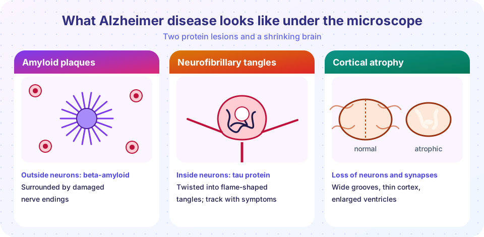
- Amyloid plaques are extracellular deposits of beta-amyloid (Aβ), a fragment cut from the amyloid precursor protein (APP). The more aggregation-prone Aβ42 form seeds plaques.
- Neurofibrillary tangles are twisted fibers of hyperphosphorylated tau protein inside neurons. Tau normally stabilizes the cell's internal tracks, and in disease it detaches and clumps. Tangles correlate better with symptoms than plaques do.
- Neuron and synapse loss causes atrophy, with widened sulci, a thin cortex and enlarged ventricles. Cholinergic neurons of the basal forebrain are lost, so acetylcholine falls.
The disease starts in the entorhinal cortex and hippocampus, which explains the early loss of new memories, and spreads to the association cortex. Most cases occur after age 65 and are multifactorial. The strongest genetic risk factor is the APOE ε4 allele. Rare inherited early-onset forms involve mutations in APP and the presenilin genes (PSEN1, PSEN2). People with Down syndrome carry three copies of the APP gene (on chromosome 21) and develop Alzheimer pathology early. The "amyloid hypothesis" has been central, and treatments that remove amyloid have had modest effects, so the role of tau, inflammation and other factors remains actively studied.
Other causes
- Vascular dementia follows strokes, including many small infarcts and white matter damage. It often occurs alongside Alzheimer disease ("mixed" dementia).
- Dementia with Lewy bodies and Parkinson disease involve alpha-synuclein protein that forms Lewy bodies in neurons. Lewy body dementia features fluctuating attention, well-formed visual hallucinations and parkinsonism. In Parkinson disease, dopamine-producing neurons of the substantia nigra are lost, which causes tremor at rest, stiffness and slowness of movement.
- Frontotemporal dementia affects the frontal and temporal lobes, causing changes in personality, behavior or language. Pathology includes tau (Pick bodies) or TDP-43 deposits.
- Huntington disease is an inherited disorder with a CAG repeat expansion in the huntingtin gene on chromosome 4, inherited in an autosomal dominant way, with atrophy of the caudate nucleus. Symptoms include involuntary movements, psychiatric problems and cognitive decline.
- Creutzfeldt-Jakob disease (CJD) is a prion disease: the normal prion protein misfolds, and the misfolded form converts others. The brain shows spongiform change, little holes in the cortex, and the disease progresses quickly. Rare forms arise from infection (variant CJD was linked to contaminated beef in the 1990s), inheritance or sporadic misfolding.
Press Run (or Ctrl + Enter) to see the result here.
Program input · what you'd type while the program runs
Type the answers before you press Run, one per line. Each time the program reads a line (with input()), it gets the next line from this box.
Common misconceptions
- "A stroke is a heart problem." A stroke is an event in the brain's blood supply, although heart disease can cause it.
- "Dementia is a normal part of aging." Some memory slowing is common with age, but dementia is a disease.
- "Plaques and tangles are the whole story." Many older people with plaques have no symptoms, and mixed pathologies are common.
- "Stroke damage is always permanent." Early treatment and rehabilitation can limit and recover function, because penumbral tissue can be saved and other brain regions can take over some tasks.
Try it
Press Run (or Ctrl + Enter) to see the console output here.
No output.
Check your understanding
Quick check · 5 questions
of 5 answered You scored / 5 · perfect!
-
1. In the brain, an infarct typically undergoes
Brain tissue liquefies after infarction and ends as a cystic cavity with gliosis. -
2. The penumbra is
The penumbra has reduced flow but is still viable. -
3. A sudden 'worst headache of life' with blood in the subarachnoid space suggests
Subarachnoid hemorrhage from a ruptured aneurysm is classic. -
4. Which protein forms intraneuronal tangles in Alzheimer disease?
Hyperphosphorylated tau forms neurofibrillary tangles. -
5. Loss of dopaminergic neurons in the substantia nigra characterizes
Parkinson disease features nigral dopamine neuron loss and Lewy bodies.
Summary
- Stroke is ischemic (thrombus, embolus, small vessel, hypoperfusion) or hemorrhagic (hypertensive, aneurysm, amyloid angiopathy).
- A brain infarct liquefies and leaves a cavity with gliosis; the penumbra is the target of early treatment.
- Alzheimer disease features amyloid plaques, tau tangles, neuron loss and atrophy beginning in the hippocampus.
- Other neurodegenerative diseases are defined by their abnormal protein or genetic cause: alpha-synuclein, huntingtin, prions.
Track your progress
Create a free account to tick off lessons and pick up where you left off on any device.
of Pathology lessons completed