


















Lesson 11 of 46 · Inflammation and Repair
Chapter 3 Overview: Inflammation and Repair
See the big picture of inflammation: what triggers it, the five classic signs, the cells and mediators involved, and how it ends in healing, scarring or chronic disease.
When you scrape your knee, the skin turns red, warms up, swells and hurts. These are not signs that something has gone wrong with your body's response; they are the response. Inflammation is the way living tissue with a blood supply reacts to injury or infection. It brings defenders to the scene, walls off the threat, clears away dead cells and prepares the ground for repair. Pathology treats it as a central topic because it is involved in nearly every disease: infections, arthritis, asthma, heart attacks, cancer and even Alzheimer disease all have an inflammatory component.
What you will learn
- What inflammation is for, and why it is double-edged.
- The five classic signs and the changes that cause them.
- The cast of cells and chemical messengers.
- The possible outcomes of an inflammatory response.
- How the lessons in chapter 3 fit together.
Key terms
Inflammation: the protective response of vascularized tissue to infection or injury.
Mediators: chemical messengers such as histamine, prostaglandins and cytokines that coordinate the response.
Leukocyte: a white blood cell: neutrophil, monocyte or macrophage, lymphocyte, eosinophil, basophil.
Exudate: protein-rich fluid that leaks from vessels into the tissue in inflammation.
Resolution: return of the tissue to normal after the cause is removed.
The big picture
Something is wrong
Sensors raise the alarm
Vessels respond
Clean up and repair
A short history
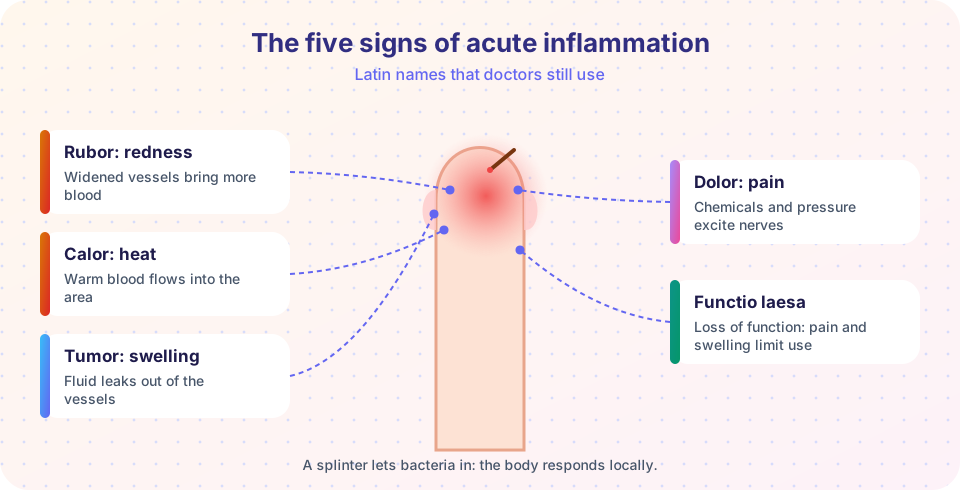
The Roman writer Celsus listed four signs of inflammation in the first century: redness, heat, swelling and pain, in Latin rubor, calor, tumor, dolor. Loss of function, functio laesa, was added later and is often credited to Rudolf Virchow. In the 1880s the Russian zoologist Élie Metchnikoff watched cells engulf particles in transparent starfish larvae and proposed that phagocytes are a defense of the body. He shared the 1908 Nobel Prize in Physiology or Medicine for work on immunity. The insight was that inflammation is not just damage: it is an active and organized defense.
What starts it
Inflammation is triggered by any harmful event: infection, tissue necrosis (from ischemia, trauma or burns), foreign material such as splinters or silica, and immune reactions such as allergy or autoimmunity. Cells detect danger through pattern recognition receptors, such as the Toll-like receptors. They recognize molecular patterns that are typical of microbes (for example bacterial cell-wall components) and molecules that are released from damaged or dying cells. Resident macrophages and mast cells then release mediators.
The cast
- Cells in the blood: neutrophils (first responders to bacteria), monocytes (they become macrophages), eosinophils (parasites and allergy), lymphocytes (the specific immune response) and platelets.
- Cells in the tissue: mast cells (histamine), macrophages, fibroblasts, and the endothelial cells that line the vessels.
- Plasma proteins: the complement system, clotting factors and antibodies, which are delivered when the vessels leak.
- Chemical mediators: histamine causes vessels to widen and leak within minutes; prostaglandins cause vessels to widen and make nerve endings more sensitive; cytokines such as TNF and interleukin-1 coordinate the response and cause fever.
From signs to mechanisms
Each cardinal sign has a mechanism.
- Redness and heat: arterioles widen, increasing blood flow to the area.
- Swelling: the smallest vessels become leaky, so protein-rich fluid escapes into the tissue.
- Pain: prostaglandins and bradykinin sensitize nerve endings, and swelling presses on them.
- Loss of function: a combination of pain, swelling and tissue damage.
At the same time, white cells attach to the vessel wall and move into the tissue. Lesson 2 of this chapter, acute inflammation, covers these steps in detail.
Systemic effects
When inflammation is strong, signals reach the whole body. Cytokines raise the temperature set-point of the brain, causing fever. The liver produces acute-phase proteins such as C-reactive protein, whose blood level is used to measure inflammation. The bone marrow releases more white cells, raising the white-cell count (leukocytosis). A patient may feel tired and lose appetite.
Outcomes
- Resolution. The injury is mild, the cause is removed, and the tissue returns to normal.
- Healing by repair and scarring. If tissue cannot regenerate or damage is great, fibrous scar replaces it.
- Abscess. Pus collects in a walled-off cavity.
- Chronic inflammation. The cause persists and the response continues, with ongoing tissue destruction and repair. See chronic inflammation.
Chapter 3 also includes healing and repair and ends with a chapter review.
Common misconceptions
- "Inflammation is the same as infection." Infection is one cause. Inflammation also happens in sterile injuries, such as a sprained ankle.
- "Inflammation is always harmful." It is a defense. Without it wounds would not heal and infections would spread. It causes harm when it is excessive, misdirected or prolonged.
- "Anti-inflammatory drugs cure disease." They reduce symptoms by blocking mediators, such as prostaglandins, but do not necessarily remove the cause.
Try it
The first block sketches how different white cells arrive at a site of bacterial injury over time. The curves are qualitative and illustrative, not measured data.
Press Run (or Ctrl + Enter) to see the result here.
Program input · what you'd type while the program runs
Type the answers before you press Run, one per line. Each time the program reads a line (with input()), it gets the next line from this box.
The second block links each cardinal sign to its main mechanism. Edit the table to quiz yourself.
Press Run (or Ctrl + Enter) to see the console output here.
No output.
Check your understanding
Quick check · 5 questions
of 5 answered You scored / 5 · perfect!
-
1. Which Latin term means swelling?
Tumor here means swelling, caused by fluid leaking from vessels. Rubor is redness, calor heat, dolor pain. -
2. What causes the redness and heat of acute inflammation?
More blood flows into the area, making it red and warm. -
3. Which cells are usually the first white cells to arrive at a bacterial injury?
Neutrophils arrive within hours; macrophages follow over the next days. -
4. Which is NOT a possible outcome of acute inflammation?
Outcomes are resolution, repair with scarring, abscess or progression to chronic inflammation. -
5. What is C-reactive protein?
CRP rises in the blood in response to cytokines such as interleukin-6.
Summary
- Inflammation is a protective, organized response of vascularized tissue to infection and injury.
- Its signs are rubor, calor, tumor, dolor and functio laesa, each explained by vessel and nerve changes.
- Resident sentinels detect danger and release mediators that bring in plasma proteins and white cells.
- Strong inflammation produces systemic effects: fever, acute-phase proteins, leukocytosis.
- Outcomes are resolution, repair with scar, abscess or chronic inflammation.
Next: Acute Inflammation
Track your progress
Create a free account to tick off lessons and pick up where you left off on any device.
of Pathology lessons completed