


















Lesson 32 of 46 · Gastrointestinal, Renal and Endocrine Pathology
Gut, liver and pancreas: ulcers, IBD, hepatitis, cirrhosis
Learn the pathology of peptic ulcers, gastric and colorectal cancer, Crohn disease, ulcerative colitis, celiac disease, hepatitis, cirrhosis, pancreatitis and pancreatic cancer.
The digestive tract is a tube lined with cells that are renewed every few days and that face acid, enzymes, bacteria and everything we swallow. That constant exposure explains most of its pathology: ulcers where the lining loses to acid, cancers where constant renewal accumulates errors, and inflammatory disease where the immune system misjudges the huge microbial population living inside. The liver and pancreas, which sit alongside, are exposed to what the gut absorbs, so they have their own versions of the same stories.
This lesson is long because it covers a lot of ground, so read it in the three parts: the tube, the liver, and the pancreas with the bile ducts.
What you will learn
- The causes and complications of peptic ulcers and the types of stomach cancer
- How Crohn disease, ulcerative colitis and celiac disease differ
- How colorectal cancer develops
- The patterns of viral, alcoholic and metabolic liver disease, and how they lead to cirrhosis
- The causes of acute pancreatitis and the pathology of pancreatic cancer
Key terms
Ulcer: a break in the lining that extends through the mucosa into deeper layers.
Transmural: involving the full thickness of the wall.
Steatosis: accumulation of fat in cells, as in fatty liver.
Portal hypertension: raised pressure in the portal vein system, usually from cirrhosis.
Jaundice: yellow discoloration from excess bilirubin.
The stomach: ulcers and cancer
Peptic ulcers form in the stomach or in the first part of the small intestine (duodenum) when the protective mucus-and-bicarbonate layer fails against acid and pepsin. The two main causes are infection with Helicobacter pylori, a curved bacterium that lives in the stomach lining, and use of nonsteroidal anti-inflammatory drugs (NSAIDs), which reduce the prostaglandins that protect the mucosa. Rarely a gastrin-secreting tumor (Zollinger-Ellison syndrome) causes extreme acid output. Barry Marshall and Robin Warren showed in the 1980s that the bacterium causes ulcers, an idea that was doubted at first, and which earned them a Nobel Prize in 2005. Ulcers are typically round and "punched out" with clean edges, and the base shows layers of necrosis, inflammation and scar. Complications are bleeding (the commonest), perforation with peritonitis, and obstruction from scarring at the stomach outlet.
Gastric adenocarcinoma is the main stomach cancer. Chronic H. pylori gastritis can progress through atrophy and intestinal metaplasia to dysplasia and cancer, a path that fits the multistep model from Chapter 5. Two forms are described. The intestinal type forms glands and follows that gastritis sequence. The diffuse type has scattered "signet ring" cells that push mucus to the side of the nucleus and infiltrates the wall, giving a thick, rigid stomach called linitis plastica. It is linked to loss of E-cadherin. Spread to a left supraclavicular lymph node is called Virchow node.
The intestines: inflammatory bowel disease and more
Inflammatory bowel disease is a pair of chronic, relapsing diseases, thought to arise from a mix of genetics, an overactive immune response to gut microbes, and the environment.
| Feature | Crohn disease | Ulcerative colitis |
|---|---|---|
| Where | Anywhere from mouth to anus; often terminal ileum and colon | Rectum and colon only |
| Pattern | Skip lesions (patches of disease between normal bowel) | Continuous from the rectum upward |
| Depth | Transmural | Mucosa and submucosa |
| Hallmarks | Cobblestone mucosa, deep fissures, non-caseating granulomas, strictures, fistulas | Crypt abscesses, pseudopolyps, no granulomas |
| Complications | Fistulas, abscesses, obstruction, malabsorption | Toxic megacolon, bleeding, higher colon cancer risk after many years |
The practical difference is that depth matters: a transmural disease creates fistulas and strictures, while a mucosal disease causes bloody diarrhea.
Celiac disease is an immune reaction to gluten (a protein in wheat, barley and rye) in genetically susceptible people, mostly carrying HLA-DQ2 or DQ8. The immune attack flattens the finger-like villi of the small bowel (villous atrophy), reducing absorption. Blood tests show antibodies to tissue transglutaminase, and the diagnosis is confirmed by biopsy.
Colorectal cancer
Most colorectal cancers are adenocarcinomas that arise from adenomatous polyps in the adenoma-carcinoma sequence introduced in Chapter 5: loss of APC, then KRAS mutation, then loss of TP53. Two inherited syndromes illustrate the genes: familial adenomatous polyposis (APC mutation; hundreds to thousands of polyps and near-certain cancer without surgery), and Lynch syndrome (mismatch-repair gene defects). Risk factors include age, family history, IBD, and a diet high in red and processed meat and low in fiber. Left-sided tumors tend to cause obstruction and bloody stool, and right-sided tumors cause slow bleeding and iron-deficiency anemia. Because polyps take years to become cancer, screening by colonoscopy or stool tests can prevent cancer by removing them.
The liver
The liver has a huge reserve and a remarkable ability to regenerate. It also receives all the blood from the gut, so toxins, drugs and microbes reach it first. The common responses to injury are inflammation (hepatitis), fat accumulation, and scarring.
Jaundice
Bilirubin is a yellow pigment made when heme from old red cells is broken down. The liver makes it water soluble (conjugated) and sends it into bile. Jaundice can come from too much made (hemolysis: unconjugated bilirubin rises), from liver cell disease (both types rise), or from blocked bile flow (obstruction: conjugated bilirubin rises, urine turns dark and stool pale). Painless jaundice in an older adult raises suspicion of a tumor at the head of the pancreas.
Viral hepatitis
Five hepatitis viruses cause most of the viral liver disease.
- Hepatitis A and E are spread by contaminated food and water, and cause acute illness but not chronic infection (E can be chronic in transplant recipients).
- Hepatitis B (a DNA virus) is spread by blood, sex and from mother to baby. It can be acute or chronic, and chronic infection leads to cirrhosis and liver cancer. Vaccination is very effective.
- Hepatitis C (an RNA virus) is spread mainly by blood. Most infections become chronic, and curative antiviral drugs now exist.
- Hepatitis D needs hepatitis B to replicate.
Injury is mostly caused by the immune system attacking infected cells, not by the virus itself. Pathology shows liver cell swelling and death, with scattered apoptotic bodies (Councilman bodies) and lymphocytes. In chronic disease the inflammation continues for months to years, and scar builds up.
Press Run (or Ctrl + Enter) to see the console output here.
No output.
Fatty and alcoholic liver disease
Fat accumulation in liver cells (steatosis) has two main causes: alcohol, and a cluster of metabolic problems linked to obesity and insulin resistance. The second is now called metabolic dysfunction-associated steatotic liver disease (MASLD), formerly "non-alcoholic fatty liver disease." Both pass through similar stages.
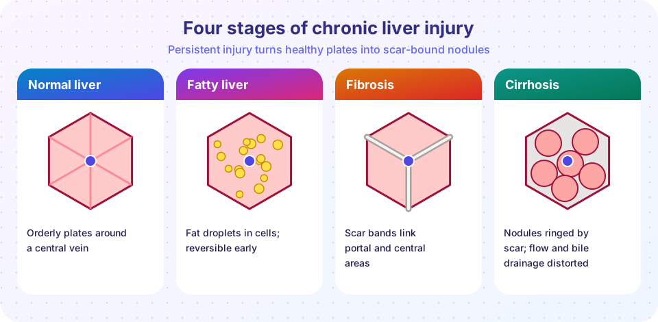
- Steatosis: fat droplets in cells; often reversible if the cause stops.
- Steatohepatitis: fat plus inflammation and cell injury, including swollen "ballooned" cells with Mallory-Denk bodies (clumped keratin) in alcoholic disease.
- Fibrosis: scar tissue spreads around the central veins and portal tracts.
- Cirrhosis: bands of scar divide the liver into regenerative nodules.
Cirrhosis
Cirrhosis is the end stage of many chronic liver diseases (alcohol, hepatitis B and C, MASLD, autoimmune and biliary disease, iron overload). It is diffuse, which means the whole liver is affected, and the nodules and scar distort blood flow. The consequences fall into two groups.
- Portal hypertension: blood backs up in the portal system. It forces open collateral veins, the most dangerous of which are esophageal varices that can bleed massively. It also causes an enlarged spleen, ascites (fluid in the abdomen), and shunting of blood that bypasses the liver.
- Liver failure: the liver cannot make albumin and clotting factors (bruising, swelling), cannot detoxify (hepatic encephalopathy, confusion from toxins such as ammonia), and cannot clear estrogen and bilirubin.
Cirrhosis also raises the risk of hepatocellular carcinoma, the most common primary liver cancer, strongly associated with chronic hepatitis B and C and cirrhosis, which may be detected by raised AFP and ultrasound screening in people at risk.
Press Run (or Ctrl + Enter) to see the result here.
Program input · what you'd type while the program runs
Type the answers before you press Run, one per line. Each time the program reads a line (with input()), it gets the next line from this box.
The pancreas and bile ducts
The pancreas has two jobs: exocrine (digestive enzymes into the duodenum) and endocrine (insulin and other hormones from the islets).
- Acute pancreatitis is autodigestion: digestive enzymes activate inside the gland and digest it. The commonest causes are gallstones blocking the common channel, and heavy alcohol use. Severe abdominal pain with high blood amylase and lipase is typical. Pathology shows fat necrosis, where released lipase breaks down fat to fatty acids that combine with calcium (chalky "soap" deposits). Severe disease can cause shock and failure of other organs.
- Chronic pancreatitis is repeated injury causing fibrosis, calcification and loss of function (maldigestion and diabetes). Alcohol is the commonest cause.
- Pancreatic ductal adenocarcinoma is one of the deadliest common cancers, in part because it is found late and spreads early. It arises from precursor lesions in small ducts and commonly carries mutations in KRAS, TP53, CDKN2A and SMAD4. Tumors in the head compress the bile duct and cause painless jaundice, while body or tail tumors present later, with pain and weight loss.
- Gallstones (most are cholesterol stones) can cause cholecystitis (inflammation of the gallbladder) or block the bile duct, causing jaundice.
Common misconceptions
- "Stress and spicy food cause ulcers." The main causes are H. pylori and NSAIDs.
- "Crohn disease and ulcerative colitis are the same." Crohn disease is transmural and patchy anywhere; ulcerative colitis is mucosal and continuous from the rectum.
- "Only heavy drinkers get cirrhosis." Hepatitis B and C, metabolic liver disease and other causes also lead to cirrhosis.
- "Jaundice is a disease." It is a sign with different causes: hemolysis, liver cell disease or blocked bile flow.
Try it
The hepatitis B table and the fibrosis model above are the main demonstrations. One more small lookup for jaundice patterns:
Press Run (or Ctrl + Enter) to see the console output here.
No output.
Check your understanding
Quick check · 5 questions
of 5 answered You scored / 5 · perfect!
-
1. Which bacterium is the main infectious cause of peptic ulcers?
H. pylori colonizes the stomach and causes chronic gastritis and ulcers. -
2. Skip lesions and non-caseating granulomas are typical of
Crohn disease is patchy and transmural, and may have granulomas. -
3. What is cirrhosis?
Cirrhosis is the end-stage pattern of diffuse fibrosis and nodule formation. -
4. Pale stool, dark urine and conjugated hyperbilirubinemia suggest
Blocked bile flow raises conjugated bilirubin, which spills into urine, while stool loses its pigment. -
5. The two most common causes of acute pancreatitis are
Gallstones and heavy alcohol use account for most cases.
Summary
- Peptic ulcers come mostly from H. pylori or NSAIDs; gastric cancer follows chronic gastritis (intestinal type) or arises as a diffuse type.
- Crohn disease is transmural, patchy and granulomatous; ulcerative colitis is mucosal, continuous and rectal; celiac disease flattens villi in response to gluten.
- Colorectal cancer typically follows the adenoma-carcinoma sequence and can be prevented by screening.
- Chronic liver injury (viral, alcohol, metabolic) leads through fibrosis to cirrhosis, with portal hypertension, liver failure and cancer risk.
- Acute pancreatitis is autodigestion; pancreatic adenocarcinoma is aggressive and often found late.
Next: Kidney disease
Track your progress
Create a free account to tick off lessons and pick up where you left off on any device.
of Pathology lessons completed