


















Lesson 30 of 46 · Cardiovascular and Respiratory Pathology
Chapter 6 review: Cardiovascular and respiratory pathology
Review plaques, infarcts, hypertension, heart failure, valve disease, COPD, fibrosis, pneumonia and lung cancer with a summary picture, table and ten-question quiz.
This chapter tied together two organs that depend on each other. If you can explain, in your own words, how a plaque causes an infarct, why a thick left ventricle fails, and what spirometry tells you about the lungs, you have the main ideas. Use the picture and table below as a checklist, then try the quiz without looking back.
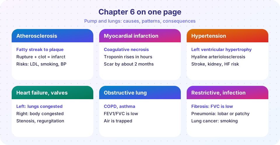
What you will learn
- How to connect each disease to its cause and consequence
- Which findings distinguish similar conditions
- Where to revisit the lessons
Key terms
Ischemia / infarction: reversible oxygen shortage / tissue death. Hypertrophy: larger cells under load. Obstructive / restrictive: slow air out / limited expansion. Consolidation: air spaces filled by exudate.
Recap table
| Disease | Mechanism | Key findings | Lesson |
|---|---|---|---|
| Atherosclerosis | Endothelial injury, LDL, foam cells, fibrous cap | Fatty streak, plaque, rupture, thrombus | Atherosclerosis |
| Myocardial infarction | Plaque rupture with occlusion | Coagulative necrosis, neutrophils at 1 to 3 d, macrophages at 3 to 7 d, scar by 2 months; troponin | Atherosclerosis |
| Hypertension | Primary (most) or secondary | Hyaline or onion-skin arterioles; LV hypertrophy | Hypertension |
| Heart failure | Pump fails; neurohormonal compensation | Left: pulmonary edema; right: systemic congestion | Hypertension |
| Valve disease | Stenosis or regurgitation | Calcific aortic, rheumatic mitral, endocarditis | Hypertension |
| COPD | Smoke, protease imbalance | Low FEV1/FVC; emphysema, chronic bronchitis | Lung disease |
| Asthma | Th2 allergic inflammation | Reversible narrowing, eosinophils, mucus plugs | Lung disease |
| Restrictive disease | Fibrosis, dust, wall disorders | Low FVC, normal or high ratio | Lung disease |
| Pneumonia | Lobar, broncho, interstitial, aspiration | Consolidation, pus, hepatization stages | Lung disease |
| Lung cancer | Smoking and other carcinogens | Adenocarcinoma, squamous, small cell | Lung disease |
Threads that connect the lessons
- Pressure and flow: narrowing of an artery lowers flow with the fourth power of radius, and a high resistance raises pressure the heart must pump against. Both ideas link atherosclerosis and hypertension.
- Compensation then decompensation: hypertrophy, neurohormonal activation and Frank-Starling are all helpful at first, and harmful if the load never lifts.
- Smoke: it injures endothelium, destroys alveolar walls, scars bronchi and causes cancer.
- Pattern recognition: many diagnoses rest on spotting a pattern (the time-dated infarct, the spirometry curve, the central small cell tumor) and tracing it back to the mechanism.
Common misconceptions
- "Angina and infarction are the same." Angina is reversible ischemia; infarction is cell death.
- "Left and right heart failure are unrelated." Left failure is the commonest cause of right failure.
- "A normal FEV1/FVC rules out lung disease." Restrictive diseases and early disease can have a normal ratio.
Questions to explain in your own words
Write a brief answer to each, then check it against the lessons.
- Why does a plaque that narrows an artery by only half sometimes cause a heart attack, while a stable narrower plaque does not?
- Explain why the weakest time for an infarcted heart wall is 3 to 7 days after the event.
- Why does left heart failure cause breathlessness while right heart failure causes ankle swelling?
- How would you distinguish an obstructive from a restrictive lung pattern using two numbers?
- Pick a lung disease and a heart disease that share a cause, and explain the shared mechanism.
Use the recap table to find where each answer lives, and reread only the part you missed.
Try it
Press Run (or Ctrl + Enter) to see the result here.
Program input · what you'd type while the program runs
Type the answers before you press Run, one per line. Each time the program reads a line (with input()), it gets the next line from this box.
Press Run (or Ctrl + Enter) to see the console output here.
No output.
Check your understanding
Quick check · 10 questions
of 10 answered You scored / 10 · perfect!
-
1. The earliest visible lesion of atherosclerosis is the
Foam cells in the intima form a fatty streak. -
2. Neutrophils dominate infarcted myocardium at about
The neutrophil infiltrate peaks at 1 to 3 days. -
3. Free-wall rupture after an infarct is most likely at
At that time macrophages have removed debris and the wall is soft and thin. -
4. Which finding points to left heart failure?
Left failure congests the lungs. -
5. Which is most likely to be secondary hypertension?
Endocrine tumors and renal disease are classic secondary causes. -
6. Rheumatic heart disease arises from
It is an autoimmune reaction by molecular mimicry. -
7. Centriacinar emphysema in the upper lobes is typical of
Smoking causes centriacinar emphysema, while alpha-1 antitrypsin deficiency causes panacinar. -
8. Which pair of results indicates a restrictive pattern?
Both volumes are reduced proportionally, so the ratio is preserved. -
9. Squamous cell carcinoma of the lung can cause hypercalcemia by secreting
Parathyroid hormone-related peptide raises blood calcium. -
10. Infective endocarditis from Staphylococcus aureus typically
S. aureus is virulent and causes acute valve destruction even on normal valves.
Summary
- Atherosclerosis causes most heart attacks through plaque rupture and thrombosis, and the infarct matures through a predictable sequence.
- Hypertension remodels vessels and the heart; heart failure congests the lungs (left) or the body (right).
- Valves narrow or leak; rheumatic, calcific and infective causes are classic.
- Spirometry separates obstructive from restrictive lung disease; pneumonia and lung cancer have recognizable patterns.
Next: Chapter 7 overview: Gastrointestinal, renal and endocrine pathology
Track your progress
Create a free account to tick off lessons and pick up where you left off on any device.
of Pathology lessons completed