


















Lesson 4 of 46 · Getting Started
How Pathologists Work: From Biopsy to Report
Follow a tissue sample from biopsy through fixation, sectioning and H&E staining to the report, and learn why test results depend on prevalence.
A biopsy is a small piece of living tissue removed so that someone can study it. It looks unremarkable in its container, but within a day or two it passes through a series of carefully designed steps that turn it into a thin, colored slice a pathologist can read. Understanding those steps is not just trivia. Every step can introduce artifacts, every stain answers a specific question, and the final report is only as reliable as the sample and the method. In this lesson you will follow a specimen from the operating room to the report and then learn the statistics that keep test results honest.
What you will learn
- The main types of sample: biopsy, resection, cytology, frozen section and autopsy.
- The steps that turn tissue into a stained slide, and what H&E shows.
- When special stains, immunohistochemistry and molecular tests are added.
- What a pathology report contains.
- How sensitivity, specificity and prevalence determine what a positive result means.
Key terms
Biopsy: removal of a small sample of tissue for examination.
Fixation: chemically preserving tissue so it stops decaying and keeps its structure; usually formalin.
H&E: hematoxylin and eosin, the routine two-dye stain.
Immunohistochemistry (IHC): staining that uses antibodies to show where a specific protein is in tissue.
Sensitivity and specificity: the fraction of diseased cases a test detects, and the fraction of healthy cases it correctly clears.
Types of specimen
Not all samples are alike, and the type decides the questions it can answer.
- Needle or punch biopsy: a tiny core or disc, enough to diagnose but not to show the whole lesion.
- Excisional biopsy and resection: the whole lump or a whole organ part. These show the edges (margins) and let the pathologist stage a cancer.
- Cytology: loose cells rather than tissue pieces. A cervical smear and fine-needle aspiration of a thyroid nodule are classic examples. They are fast and minimally invasive, but the tissue architecture is lost.
- Frozen section: during an operation, a piece is frozen, cut and stained within minutes so the surgeon can adapt the operation. It is quicker but less detailed than the standard method.
- Autopsy: examination of a body after death to find the cause of death or to answer clinical questions.
From tissue to slide
Almost all routine histology uses the same sequence.
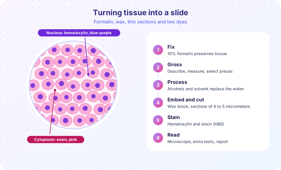
- Fixation. The specimen is placed in neutral buffered formalin (a dilute solution of formaldehyde). Formaldehyde links proteins together, stops enzymes from digesting the tissue and kills microbes. Without it, cells would digest themselves within hours. Penetration is slow, so large specimens are cut open and fixed for many hours.
- Grossing. The pathologist (or an assistant) describes the specimen: size, weight, color, texture, margins. Representative pieces are chosen and placed in small cassettes. This is a sampling step: whatever is not submitted cannot be examined.
- Processing. Water in the tissue is replaced stepwise by alcohol, then a clearing solvent, then molten paraffin wax.
- Embedding and sectioning. The tissue is set in a wax block, and a microtome cuts sections that are only a few micrometers thick (a typical section is about 4 micrometers, thinner than most single cells are wide, so a slide shows a thin slice of cells, not whole ones).
- Staining. The wax is dissolved, and the section is stained, most often with H&E.
- Reading. A pathologist studies the slide at several magnifications, correlates it with the clinical information and writes the report.
Why H&E works
Hematoxylin, used with a metal salt that acts as a mordant, behaves like a basic dye. It binds to acidic components, mainly the DNA and RNA in nuclei, and turns them blue to purple. Eosin is an acidic dye that binds to basic components such as most cytoplasmic proteins and collagen, turning them shades of pink. That is why the schematic above shows purple nuclei in pink cells. The pair is cheap, robust and shows structure, cell shape and nuclear detail in a single glance, so it has been the standard for more than a century. Learn to ask of every slide: what is the architecture, what do the cells look like, what is the nucleus doing, what is the background?
When H&E is not enough
When H&E leaves a question open, the pathologist orders extra tests on the same block or slides.
| Tool | What it shows | Example question |
|---|---|---|
| Special stains | A chemical component in tissue | Is there collagen scar (trichrome, blue or green)? Are there fungi (PAS, magenta)? Acid-fast bacteria (Ziehl-Neelsen)? Iron (Prussian blue)? |
| Immunohistochemistry | A specific protein, using a labelled antibody | Is this tumor from breast tissue? Does it carry the HER2 or the estrogen receptor protein? How many cells are dividing (Ki-67)? |
| Molecular tests | DNA, RNA or chromosome changes | Does the tumor carry a mutation that predicts response to a particular drug? |
| Culture and PCR | Living microbes or their genetic material | Which organism caused this infection? |
Each of these is a way to turn a visual impression into a testable answer.
The pathology report
A typical surgical pathology report has these parts: patient and specimen identifiers; clinical history provided by the clinician; the gross description; the microscopic description; the final diagnosis; and notes on ancillary tests. For cancers, a synoptic report lists key items in a standard checklist: tumor type, grade, size, margin status, spread to lymph nodes. Standardizing the checklist means that nothing important is forgotten and different hospitals report in the same way.
Reports carry uncertainty. Pathologists sometimes agree to disagree: on borderline lesions, even experienced readers can differ. That is why difficult or high-stakes cases are often shown to a second pathologist or discussed at a multidisciplinary meeting.
Sensitivity, specificity and what a positive result means
Every diagnostic test makes two kinds of mistake. A false negative misses a real case. A false positive flags a healthy person.
- Sensitivity = true positives divided by all people who truly have the disease.
- Specificity = true negatives divided by all people who truly do not.
These describe the test. What a patient wants to know is different: "given a positive result, what is the probability that I have the disease?" That probability is the positive predictive value (PPV), and it depends on how common the disease is (the prevalence). Using Bayes' rule,
PPV = (sensitivity × prevalence) / (sensitivity × prevalence + (1 - specificity) × (1 - prevalence))
Suppose a screening test has 95 percent sensitivity and 90 percent specificity. If the disease affects 1 percent of the people tested, then among 1,000 people there are 10 true cases. The test detects 9.5 of them. Of the 990 healthy people, 10 percent (99) are falsely flagged. So only about 9.5 of 108.5 positives are real: a PPV of roughly 9 percent. If the same test is used in a high-risk group where prevalence is 30 percent, the PPV climbs to about 80 percent. The test did not change; the population did. This is why doctors confirm a positive screening test with a more definitive test such as a biopsy.
Common misconceptions
- "A positive test means I have the disease." Not necessarily. Whether a positive is likely to be real depends on prevalence as well as on the test.
- "A negative slide means no disease." A small biopsy can miss a lesion (sampling error). A negative result lowers probability, it does not always rule it out.
- "The pathologist sees the whole organ." A slide shows a thin slice of selected pieces. Good grossing and good clinical information matter.
Try it
The first block plots the positive predictive value for a test with fixed sensitivity and specificity as prevalence changes. The numbers are illustrative, not from a real test.
Press Run (or Ctrl + Enter) to see the result here.
Program input · what you'd type while the program runs
Type the answers before you press Run, one per line. Each time the program reads a line (with input()), it gets the next line from this box.
The second block is a small stain finder: describe what you want to show and it names a typical choice. It is a study aid with the common textbook pairings.
Press Run (or Ctrl + Enter) to see the console output here.
No output.
Check your understanding
Quick check · 5 questions
of 5 answered You scored / 5 · perfect!
-
1. What is the main purpose of fixing tissue in formalin?
Formalin cross-links proteins, halts self-digestion and kills microbes, so the structure can be studied. -
2. In H&E staining, which structure is usually blue-purple?
Hematoxylin binds acidic nucleic acids, so nuclei look blue to purple. Eosin colors most cytoplasm and collagen pink. -
3. A surgeon needs an answer during an operation. Which method is suited to this?
A frozen section can be cut and stained within minutes to guide the operation. -
4. A test has 95% sensitivity and 90% specificity. Why is its positive predictive value low when only 1% of those tested have the disease?
At low prevalence, even a small false-positive rate among many healthy people outnumbers the true positives. -
5. Which tool would best show where a specific protein is located in a tissue section?
IHC uses a labelled antibody that binds the protein of interest.
Summary
- Samples range from tiny biopsies and cytology to resections, frozen sections and autopsies.
- Routine histology: fix, gross, process, embed, cut, stain, read.
- H&E shows nuclei blue-purple and cytoplasm and collagen pink; special stains, IHC and molecular tests answer specific questions.
- A pathology report combines gross and microscopic findings and, for cancer, a standard checklist.
- Sensitivity and specificity describe a test, but the meaning of a positive result also depends on prevalence.
Next: Chapter 1 Review
Track your progress
Create a free account to tick off lessons and pick up where you left off on any device.
of Pathology lessons completed