


















Lesson 24 of 46 · Neoplasia
Grading, staging and diagnosis of cancer
See how pathologists diagnose a tumor, grade how abnormal it looks, stage how far it has spread with TNM, and why tumor markers need careful interpretation.
When a lump is found, the first question is "what is it?" and the second is "how dangerous is it?" Pathology answers the first by looking at tissue and the second by describing the tumor with two scales that sound alike but measure different things. Grade describes the cells: how abnormal they look. Stage describes the patient's disease: how far it has spread. Together with the type of tumor, they form the language that surgeons, oncologists and radiologists share.
This lesson follows a specimen from biopsy to report, then examines tumor markers, which are often misunderstood. As with all of this course, it is education, not advice about any individual case.
What you will learn
- How tissue is sampled and what the pathologist reports
- What tumor grade means and how it is assigned
- How the TNM system produces a stage
- Why tumor markers help monitoring more than diagnosis
- What paraneoplastic syndromes and cachexia are
Key terms
Biopsy: removal of cells or tissue for microscopic examination.
Grade: a measure of how abnormal tumor cells appear (differentiation, nuclear atypia, mitoses).
Stage: the extent of disease, often by the TNM system.
Tumor marker: a substance (often a blood protein) made by or in response to a tumor.
Immunohistochemistry (IHC): using labeled antibodies to detect proteins in tissue.
From lump to diagnosis
Symptoms, examination and imaging (X-ray, ultrasound, CT, MRI) can suggest a tumor, but they rarely prove what it is. The diagnosis is made by examining tissue or cells under the microscope. The choice of sample depends on the site:
- Cytology looks at individual cells. A Pap smear from the cervix and fine-needle aspiration (FNA) of a thyroid nodule or lymph node are examples. It is fast and minimally invasive, but it shows little architecture.
- Core needle biopsy removes a thin cylinder of tissue, preserving structure. It is common for breast, prostate and liver.
- Endoscopic biopsy takes small samples from the lining of the gut or airways.
- Incisional or excisional biopsy removes part or all of a lesion. A mole suspected of melanoma is removed whole rather than sampled, so that its depth can be measured.
- Frozen section gives a quick answer during surgery, for example to check whether a margin is clear. It is less detailed than permanent sections.
In the lab the tissue is fixed in formalin, embedded in paraffin, cut into slices about 4 micrometers thick, and stained with hematoxylin and eosin (H&E), which colors nuclei blue-purple and cytoplasm pink. A pathologist then asks: Is it neoplastic? Benign or malignant? What type? What grade? Has it been completely removed (are the margins clear)? Are there cancer cells in vessels or nerves?
Special stains and molecular tests
H&E often cannot say where a poorly differentiated tumor came from. Immunohistochemistry uses antibodies that stain proteins characteristic of a cell lineage. Cytokeratins point to carcinoma, vimentin to mesenchymal tumors, CD45 to lymphoid cells, and S-100 to melanoma and nerve-sheath tumors, for example. In breast cancer, IHC for the estrogen receptor, progesterone receptor and HER2 guides treatment. Increasingly, molecular tests look for specific mutations, such as EGFR in some lung cancers, BRAF in melanoma, or KRAS in colon cancer, and flow cytometry and cytogenetics are central to diagnosing leukemias and lymphomas.
Grading: how abnormal do the cells look?
Grade is assigned by the pathologist and refers to the tumor's cells. In general it reflects the degree of differentiation and the number of dividing cells:
| Grade | Typical meaning |
|---|---|
| G1 | Well differentiated; cells resemble normal tissue |
| G2 | Moderately differentiated |
| G3 | Poorly differentiated; little resemblance |
| G4 | Undifferentiated or anaplastic (not used for all sites) |
Different organs have their own systems. In breast cancer the Nottingham system scores gland formation, nuclear size and shape, and the mitotic count. In prostate cancer the Gleason system, now reported as five Grade Groups, looks at the glandular architecture. Some centers also report the Ki-67 index, the proportion of cells staining for a marker of proliferation.
Grade matters because a higher-grade tumor tends to grow faster and spread earlier. But grade is not destiny. A low-grade tumor that has been left untreated for years can still spread, and a high-grade tumor caught early can be cured.
Staging: how far has it spread?
The most widely used staging system is TNM, maintained jointly by the American Joint Committee on Cancer (AJCC) and the Union for International Cancer Control (UICC). It asks three questions.
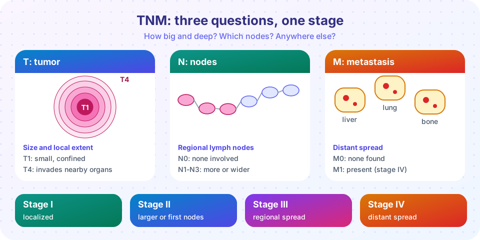
- T (tumor): the size or local extent of the primary tumor. Tis is carcinoma in situ, and T1 to T4 describe increasing size or depth of invasion, with T4 usually meaning invasion of neighboring structures.
- N (nodes): whether cancer cells are in regional lymph nodes. N0 is none; N1 to N3 describe increasing numbers or locations of involved nodes.
- M (metastasis): M0 means no distant metastasis; M1 means distant spread.
The three letters are combined into an overall stage group, usually Stage 0 (in situ) to Stage IV. Stage IV generally means distant metastasis. The exact rules differ by organ: the criteria for T1 in the breast have nothing to do with T1 in the colon, so you must never compare numbers across cancers.
Two kinds of staging exist. Clinical stage (cTNM) is based on examination and imaging before treatment. Pathologic stage (pTNM) is based on the specimen removed at surgery and is more accurate. Staging is often a stronger predictor of outcome than grade, and it drives the treatment plan: early-stage tumors may be treated by surgery alone, while spread beyond the original site usually calls for systemic therapy.
Many hospitals write the sentence in a pathology report in a fixed way: type, grade, size, margins, lymphovascular invasion, nodal status, and pTNM. Reading a report in that order makes it much easier.
Press Run (or Ctrl + Enter) to see the console output here.
No output.
Tumor markers: useful, but not a test for cancer
A tumor marker is a substance in blood, urine or tissue that is raised in some cancers. Examples:
| Marker | Associated with | Also raised in |
|---|---|---|
| PSA | Prostate cancer | Benign prostate enlargement, prostatitis |
| CEA | Colorectal and other adenocarcinomas | Smoking, inflammation |
| AFP | Liver cancer, some germ cell tumors | Cirrhosis, pregnancy |
| hCG | Germ cell tumors and trophoblastic tumors | Pregnancy |
| CA-125 | Ovarian cancer | Endometriosis, menstruation |
| Calcitonin | Medullary thyroid carcinoma | Some other conditions |
No marker is both highly sensitive and highly specific for early cancer, so most are used to monitor known disease (does a level fall after surgery? does it rise again?) rather than to screen. The reason is mathematical, not just biological. When a disease is rare, most positive results come from people who do not have it, even when the test is good. This is a consequence of Bayes' theorem.
Try it
Change the sensitivity, specificity or prevalence and rerun. The marker here is hypothetical.
Press Run (or Ctrl + Enter) to see the result here.
Program input · what you'd type while the program runs
Type the answers before you press Run, one per line. Each time the program reads a line (with input()), it gets the next line from this box.
The same marker is more useful in a group where the disease is common (for example, patients already known to have a tumor) than in the general public.
When cancer acts at a distance
Tumors harm the body by direct effects (pressure, obstruction, bleeding) and by systemic effects.
- Paraneoplastic syndromes are effects not caused by the tumor mass or spread but by substances the tumor makes. Small cell lung cancer can secrete ACTH (causing Cushing syndrome) or ADH (causing low sodium, SIADH). Squamous cell carcinomas can produce parathyroid hormone-related peptide, causing high blood calcium. Pancreatic cancer is linked to migratory venous thrombosis.
- Cachexia is a wasting syndrome of weight loss, muscle loss and weakness that is not caused by poor eating alone. Cytokines such as TNF help drive it, and it contributes to a large share of cancer deaths.
These syndromes can be the first sign of a hidden tumor, so recognizing them is a point of clinical value.
Common misconceptions
- "Grade and stage are the same." Grade describes the cells; stage describes the spread. A tumor can be high grade and early stage, or low grade and late stage.
- "A normal tumor marker rules out cancer." Many early cancers do not raise the marker, and some markers are normal in a large number of patients with the disease.
- "A biopsy can spread cancer." Properly performed biopsies carry very small risks of this; the benefits of an accurate diagnosis outweigh them.
Check your understanding
Quick check · 5 questions
of 5 answered You scored / 5 · perfect!
-
1. Which describes tumor grade?
Grade is about the cells' appearance (differentiation and proliferation); stage is about spread. -
2. In TNM, M1 means
M0 is no distant metastasis; M1 means distant spread, which usually corresponds to stage IV. -
3. Why is PSA a poor stand-alone diagnostic test for prostate cancer?
PSA is made by normal prostate too, and benign enlargement or inflammation raises it. -
4. Which technique stains proteins in tissue with labeled antibodies?
IHC uses antibodies against proteins that identify cell lineage or guide therapy. -
5. A tumor secreting ACTH and causing Cushing syndrome is an example of
Hormone effects caused by substances the tumor produces are paraneoplastic syndromes.
Summary
- Diagnosis rests on tissue: cytology, needle or surgical biopsy, with H&E, IHC and molecular tests.
- Grade describes the cells; stage describes how far the cancer has spread.
- TNM combines tumor extent, lymph nodes and distant metastasis into a stage group whose rules differ for each organ.
- Tumor markers mostly help monitor disease; low prevalence makes false positives common.
- Paraneoplastic syndromes and cachexia are effects of tumors at a distance.
Next: Chapter review
Track your progress
Create a free account to tick off lessons and pick up where you left off on any device.
of Pathology lessons completed