


















Lesson 25 of 46 · Neoplasia
Chapter 5 review: Neoplasia
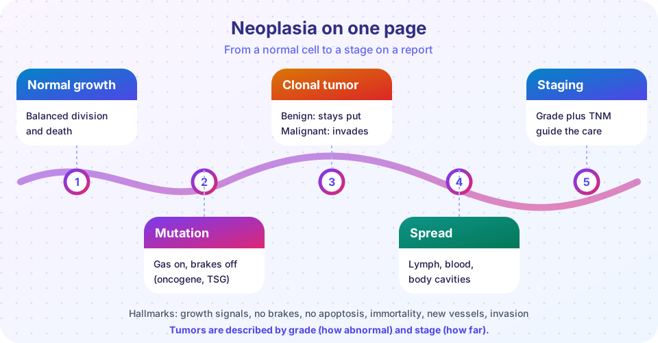
Review neoplasia in one picture: names, benign versus malignant, oncogenes and suppressors, grading and staging, with a recap table and a ten-question quiz.
You have now followed a tumor from a single mutated cell to a stage on a pathology report. This page gathers the chapter's ideas into one picture, a recap table and a longer self-test. If any row of the table feels shaky, follow the link back to the lesson before moving on to the cardiovascular and respiratory chapter.
What you will learn
- How the four lessons of the chapter connect
- Which facts are most often tested or most useful at the bedside
- Where your understanding still has gaps
Key terms
Neoplasm: autonomous new growth. Oncogene: gain-of-function growth gene. Tumor suppressor: loss-of-function brake gene. Grade: cell abnormality. Stage: extent of spread.
Recap table
| Topic | Key facts | Lesson |
|---|---|---|
| Naming | -oma = benign; carcinoma = malignant epithelium; sarcoma = malignant mesenchyme; melanoma, lymphoma, mesothelioma and seminoma are malignant | Names and growth |
| Benign vs malignant | Differentiation, growth rate, invasion, metastasis; metastasis is the most reliable sign | Names and growth |
| Precancer | Hyperplasia and metaplasia are reversible; dysplasia can progress; in situ means basement membrane intact | Names and growth |
| Spread | Invasion, intravasation, survival, arrest and exit, colonization; lymphatic, blood and cavity routes | Names and growth |
| Genes | Oncogenes (RAS, MYC, BCR-ABL, HER2) one hit; suppressors (TP53, RB1, APC, BRCA) two hits | Carcinogenesis |
| Causes | Chemicals, radiation, viruses (HPV, HBV, HCV, EBV), inflammation, heredity | Carcinogenesis |
| Hallmarks | Growth signals, no brakes, no apoptosis, telomerase, angiogenesis, invasion, metabolism, immune evasion | Carcinogenesis |
| Diagnosis | Tissue is the gold standard; cytology, biopsy, IHC, molecular tests | Grading, staging and diagnosis |
| Grade and stage | Grade = cells; stage = spread (TNM) | Grading, staging and diagnosis |
| Markers | PSA, CEA, AFP, hCG, CA-125: mostly for monitoring | Grading, staging and diagnosis |
Linking the ideas
A few cross-links make the chapter easier to remember.
- Why in situ is curable: a lesion confined by the basement membrane has not reached the vessels that carry cells away, so cure is often possible by removing it. That is the logic of screening.
- Why age matters: each step of multistep carcinogenesis is rare, and the chance that one lineage accumulates all of them rises with years. Inherited first hits shorten the wait.
- Why stage drives treatment: stage tells you whether the problem is local (surgery, radiation) or systemic (drugs), and grade fine-tunes how aggressive treatment should be.
- Why targeted drugs exist: because the same few broken proteins recur (BCR-ABL, HER2, EGFR, BRAF), a drug that blocks one can be used in everyone whose tumor carries it.
Common misconceptions
- "Cancer is a single disease." It is hundreds of diseases that share a set of capabilities.
- "A benign tumor can never turn malignant." Some benign neoplasms, such as colon adenomas, are premalignant, although most never progress.
- "Stage IV is always untreatable." Stage IV is the most advanced category, but treatment can control disease for years in many cancers.
Questions to explain in your own words
Close the lesson and write a short answer to each prompt, then compare with the text.
- Why does a carcinoma in situ have a much better outlook than an invasive carcinoma of the same organ?
- How do an oncogene and a tumor suppressor differ in the number of hits needed, and why?
- What is the difference between grade and stage, and which one tends to decide whether the cancer has left its organ?
- Why do many false alarms appear when a marker is used to screen for a rare cancer?
- Choose one cancer and trace it from a normal cell to a stage on a report, mentioning at least one cause, one gene change and one sign under the microscope.
If you cannot answer one of these, go back to the lesson linked in the table and reread only that part.
Try it
Press Run (or Ctrl + Enter) to see the console output here.
No output.
Press Run (or Ctrl + Enter) to see the result here.
Program input · what you'd type while the program runs
Type the answers before you press Run, one per line. Each time the program reads a line (with input()), it gets the next line from this box.
Check your understanding
Quick check · 10 questions
of 10 answered You scored / 10 · perfect!
-
1. Which tumor is malignant?
Lymphoma is one of the malignant tumors with an -oma ending. The others are benign. -
2. A lesion where abnormal cells fill the whole epithelium but the basement membrane is intact is
Full-thickness dysplasia without breach of the basement membrane is carcinoma in situ. -
3. Which gene change typifies an oncogene?
Oncogenes arise from gain-of-function changes; the others are loss-of-function changes in tumor suppressors. -
4. In Knudson's retinoblastoma model, the inherited form needs how many additional somatic hits?
One hit is inherited; a single further hit in a retinal cell is enough. -
5. Which is not a hallmark of cancer?
Cancer cells are typically less differentiated, not more; the other three are hallmarks. -
6. Carcinomas usually spread first via
Lymphatic spread to regional nodes is typical for carcinomas. -
7. A pathologist reports 'poorly differentiated, G3'. This describes
G1 to G3 describe grade, how abnormal the cells appear. -
8. Why are tumor markers rarely used alone to screen for cancer?
Low sensitivity and specificity plus low prevalence give poor predictive value. -
9. Which virus protein pair inactivates p53 and RB?
High-risk HPV E6 promotes p53 breakdown and E7 inactivates RB. -
10. Stage IV in TNM generally implies
M1 disease is usually grouped as stage IV.
Summary
- Names tell you tissue of origin and behavior, with a few famous exceptions.
- Benign tumors stay local; malignant ones invade and metastasize through a multistep cascade.
- Oncogenes act dominantly and suppressors recessively; cancer needs several changes.
- Grade is about the cells, stage about spread, and tissue is the gold standard for diagnosis.
Next: Chapter 6 overview: Cardiovascular and respiratory pathology
Track your progress
Create a free account to tick off lessons and pick up where you left off on any device.
of Pathology lessons completed