


















Lesson 39 of 46 · Infectious and Neurological Disease
CNS infection, demyelination and brain tumors
Learn the pathology of meningitis and encephalitis, multiple sclerosis and other demyelinating diseases, brain tumors, raised intracranial pressure and herniation.
The central nervous system (CNS) is protected by the skull, the meninges and the blood-brain barrier, a tight lining of capillaries that keeps most blood-borne substances out. These protections are why infection of the brain is relatively rare, and why, when it happens, it is serious: the immune system has limited access, and swelling in a closed skull quickly becomes a threat to life.
This lesson covers three families of brain disease: infection (meningitis, encephalitis, abscess), immune attack on myelin (multiple sclerosis), and tumors. It ends with the physics that unites them, the pressure inside the skull.
What you will learn
- The organisms and cerebrospinal fluid findings of bacterial and viral meningitis
- The patterns of encephalitis and brain abscess
- The pathology of multiple sclerosis
- The main brain tumors and how they behave
- Why raised intracranial pressure and herniation are dangerous
Key terms
Meninges: the three membranes (dura, arachnoid, pia) covering the brain and spinal cord.
Cerebrospinal fluid (CSF): the clear fluid that cushions the brain and fills the ventricles and the subarachnoid space.
Myelin: the fatty insulation around axons that speeds conduction; made by oligodendrocytes in the CNS.
Glioma: a tumor of glial cells.
Herniation: displacement of brain tissue across a rigid boundary because of pressure.
Infections of the nervous system
Meningitis
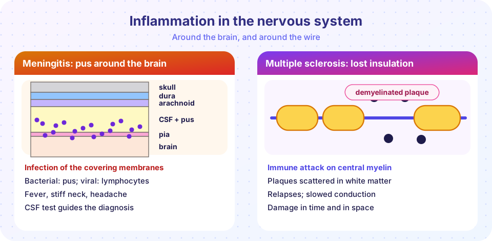
Meningitis is inflammation of the meninges, usually caused by infection. Classic symptoms are fever, headache, a stiff neck and sensitivity to light, sometimes with a rash and confusion. Bacterial meningitis is an emergency. A lumbar puncture samples the CSF, which gives the diagnosis.
The likely bacterium depends on age:
- Newborns: group B Streptococcus and Escherichia coli.
- Children and adults: Streptococcus pneumoniae and Neisseria meningitidis (the meningococcus, which can also cause a purpuric rash and sepsis). Haemophilus influenzae type b was once common in children and has become rare in countries with vaccination.
- Older adults and the immunocompromised: Listeria monocytogenes, in addition to the above.
The brain surface is coated with pus, and neutrophils fill the subarachnoid space. Complications include blocked CSF drainage (hydrocephalus), cranial nerve injury such as deafness, inflamed vessels causing infarcts, and brain swelling. Viral meningitis (often enteroviruses) is usually milder and self-limited.
Press Run (or Ctrl + Enter) to see the console output here.
No output.
Encephalitis and abscess
Encephalitis is inflammation of the brain itself, usually viral. Herpes simplex virus type 1 is the most important cause in developed countries; it infects the temporal lobes and causes hemorrhagic necrosis, with intranuclear inclusions in neurons. Arboviruses (mosquito-borne viruses such as West Nile virus) cause seasonal outbreaks. Rabies travels from the site of a bite along nerves to the brain, producing Negri bodies in neurons and an almost always fatal illness if not prevented. In immunosuppressed patients, JC virus causes progressive multifocal leukoencephalopathy by destroying oligodendrocytes, and Toxoplasma and Cryptococcus cause mass lesions and meningitis.
A brain abscess is a collection of pus within the brain tissue, usually from infection spread from the ears, sinuses or teeth, from the bloodstream (for example from endocarditis) or from head trauma. On a brain scan it appears as a ring with a dark center. Brain abscesses are a mass lesion, so they cause headache, focal deficits and raised pressure as well as fever.
Demyelinating disease: multiple sclerosis
Multiple sclerosis (MS) is the commonest demyelinating disease of the CNS and a leading cause of neurological disability in young adults, affecting women more than men. It is an autoimmune disease in which T cells (and B cells) attack myelin proteins. Genetic susceptibility (certain HLA variants), environmental factors (infection with Epstein-Barr virus, low vitamin D, smoking) and geography all play a part.
Pathology. The hallmark is the plaque: a sharply bordered area of white matter in which myelin is lost and axons are relatively preserved, at least early. Plaques are often located around the ventricles, in the optic nerves and in the brainstem and spinal cord. Active plaques show lymphocytes and macrophages containing myelin debris around small veins. Chronic plaques show a scar of astrocytes (gliosis) and loss of oligodendrocytes. As axons are also lost over time, disability builds.
Clinical features. MS is "disseminated in time and space": different symptoms occur in different places and at different times. Common features are optic neuritis (painful visual loss), weakness, numbness, a disturbance of balance and eye movements, and bladder problems. The most common course is relapsing-remitting, with attacks followed by recovery, which can later become progressive. Diagnosis combines the clinical story, MRI, and CSF testing, which often shows oligoclonal bands (antibodies produced inside the CNS).
Related conditions include neuromyelitis optica (antibodies to aquaporin-4 on astrocytes), acute disseminated encephalomyelitis (often after infection or vaccination), and, in the peripheral nervous system, Guillain-Barré syndrome, a demyelinating disease that follows infections such as Campylobacter and causes ascending weakness.
Brain tumors
Tumors of the brain are either primary (arising in the brain or its coverings) or metastatic. In adults, metastases are the most common brain tumors: they usually come from lung, breast, melanoma, kidney and colon cancers, are often multiple, and sit at the border between gray and white matter. Primary brain tumors are classified by the cell of origin and, increasingly, by their genetic changes.
| Tumor | Cell of origin | Key features |
|---|---|---|
| Glioblastoma | Astrocyte | The most common malignant primary tumor in adults; necrosis with "pseudopalisading" cells and abnormal vessel growth; may cross the corpus callosum; very poor outlook |
| Diffuse astrocytoma / oligodendroglioma | Glial cells | IDH-mutant; oligodendroglioma has 1p/19q codeletion, "fried-egg" cells, calcification; better outlook |
| Meningioma | Arachnoid cells | Outside the brain tissue, usually benign, whorls and psammoma bodies; more common in women; NF2 |
| Schwannoma | Schwann cells | Often at the vestibular nerve (hearing loss), NF2, Antoni A and B patterns |
| Medulloblastoma | Primitive cerebellar cells | Child's tumor of the cerebellum, small blue cells, Homer Wright rosettes |
| Ependymoma | Ependymal cells | Lines the ventricles; may block CSF flow |
| Pituitary adenoma | Pituitary cells | Hormone excess or mass effect on vision |
Brain tumors almost never metastasize outside the CNS, but they are dangerous because they occupy space and invade vital tissue. Because tumor grade and genetics (for example the IDH mutation) strongly influence prognosis, current classification systems combine appearance with molecular testing.
Pressure inside the skull
The skull is a closed box holding three things: brain, blood and CSF. The Monro-Kellie doctrine says that the total volume is fixed, so if one component grows (a tumor, a hemorrhage, swelling after a stroke, an abscess), another must shrink or pressure rises. At first, CSF and venous blood are squeezed out to compensate, but once that reserve is spent, small increases in volume cause large increases in pressure.
Press Run (or Ctrl + Enter) to see the result here.
Program input · what you'd type while the program runs
Type the answers before you press Run, one per line. Each time the program reads a line (with input()), it gets the next line from this box.
When pressure rises, brain tissue can be pushed across the dural folds and through the openings of the skull. This is herniation.
- Subfalcine herniation: the cingulate gyrus is pushed under the falx cerebri.
- Transtentorial (uncal) herniation: the medial temporal lobe is pushed over the edge of the tentorium and presses on the third cranial nerve (a dilated pupil on that side) and the brainstem.
- Tonsillar herniation: the cerebellar tonsils are pushed through the foramen magnum and compress the brainstem centers that control breathing and heart rate, which is rapidly fatal.
Warning signs of raised pressure include headache, vomiting, drowsiness and swelling of the optic disc. Doctors treat the cause, and may reduce brain swelling.
Common misconceptions
- "Meningitis and encephalitis are the same." Meningitis affects the coverings, encephalitis the brain tissue; the two can coexist.
- "Multiple sclerosis is a muscle disease." It affects the CNS's myelin, not muscle.
- "A brain tumor is always cancer that started in the brain." Metastases from other organs are the most common tumors in the adult brain.
- "Brain tumors spread through the body." Primary brain tumors very rarely do; they harm by local invasion and pressure.
Try it
Press Run (or Ctrl + Enter) to see the result here.
Program input · what you'd type while the program runs
Type the answers before you press Run, one per line. Each time the program reads a line (with input()), it gets the next line from this box.
Check your understanding
Quick check · 5 questions
of 5 answered You scored / 5 · perfect!
-
1. Which CSF pattern is typical of bacterial meningitis?
Bacteria attract neutrophils and consume glucose. -
2. The virus that most commonly causes severe focal encephalitis of the temporal lobes is
HSV-1 causes hemorrhagic necrosis of the temporal and orbitofrontal regions. -
3. In multiple sclerosis, plaques consist of
MS plaques are demyelinated areas in white matter with inflammation and gliosis. -
4. The most common brain tumors in adults overall are
Metastases, especially from lung and breast, outnumber primary brain tumors. -
5. Which herniation can compress the brainstem respiratory centers?
Cerebellar tonsils pushed through the foramen magnum compress the medulla.
Summary
- Meningitis is inflammation of the meninges (bacterial: pus and low glucose; viral: lymphocytes); encephalitis involves brain tissue, with HSV-1 as the key example.
- Multiple sclerosis is an autoimmune demyelinating disease with plaques disseminated in space and time.
- Brain tumors include glioblastoma, meningioma, schwannoma and medulloblastoma, while metastases are the most common in adults.
- The rigid skull obeys the Monro-Kellie doctrine, and raised pressure leads to herniation.
Next: Chapter review
Track your progress
Create a free account to tick off lessons and pick up where you left off on any device.
of Pathology lessons completed