


















Lesson 41 of 46 · Projects and Practice
Practice: case reasoning from findings to diagnosis
Practice the pathologist's five-step method on four fictional cases drawn from chapters 5 to 8, with a reveal widget, worked reasoning and a self-test.
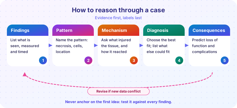
You have learned a lot of facts. Pathologists, however, rarely get asked for facts; they get handed a set of findings and asked what is going on. This lesson practices that skill using the five steps from the picture below: list the findings, name the pattern, ask about mechanism, choose the diagnosis, and predict the consequences. It uses four fictional cases, built from material in chapters 5 to 8.
Educational information only. The cases are invented teaching vignettes, not real patients, and they simplify real medicine. They train reasoning, not diagnosis or treatment decisions.
What you will learn
- A repeatable five-step method for pathology cases
- How to separate "what we see" from "what it means"
- How to use a differential diagnosis and keep it open
- How to link a diagnosis to its complications
Key terms
Differential diagnosis: the list of conditions that could explain the findings.
Pathognomonic: a finding so characteristic that it nearly proves a diagnosis (these are rarer than people think).
Anchoring: sticking to the first idea and ignoring evidence against it.
Paraneoplastic: an effect of a tumor caused by substances it releases, not by its mass.
The five steps
- Findings
- Pattern
- Mechanism
- Diagnosis
- Consequences
A worked example
Here is a case, reasoned out loud, before you try the others.
A 62-year-old man dies suddenly six days after being treated for a heart attack. At autopsy, the pericardial sac is full of blood. The front wall of the left ventricle contains a soft, yellow-tan area with a small tear. Under the microscope the area contains dead muscle, many macrophages and no scar yet.
- Findings. Death at six days after an infarct; blood in the pericardial sac; a soft, yellow area with a tear; macrophages; no scar.
- Pattern. Dead muscle plus macrophages and no scar fits the 3-to-7-day window of an infarct (Chapter 6): macrophages have cleaned up debris and the wall is soft and thin.
- Mechanism. An infarct from coronary occlusion. At that stage the wall is at its weakest.
- Diagnosis. Rupture of the left ventricular free wall, with hemopericardium and cardiac tamponade. A competing idea, a ruptured aortic dissection, would not explain the soft yellow infarct with macrophages.
- Consequences. Blood in the pericardial sac stops the heart from filling, which leads to collapse in minutes.
Notice how the timing (six days) and the cell type (macrophages) did most of the work. This is typical: a small number of well-chosen features decide the diagnosis.
A second worked example: when the diagnosis is not obvious
Not every case ends in a single textbook answer. Consider this one.
A 71-year-old woman has been increasingly forgetful for two years. Her family says the decline has come in steps, with a sudden worsening after a brief episode of slurred speech a year ago. She has long-standing high blood pressure. A brain scan shows several small old infarcts in the deep white matter and also shrinkage of the hippocampus.
- Findings. A two-year decline with step-like worsening; one transient episode of slurred speech; hypertension; small deep infarcts; hippocampal atrophy.
- Pattern. Small deep infarcts are the lacunes of small vessel disease (Chapter 8). Hippocampal shrinkage is the early site of Alzheimer pathology.
- Mechanism. Long-standing hypertension injures small penetrating arteries and produces lacunar infarcts and white matter damage. A separate neurodegenerative process may also be present.
- Diagnosis. Probably mixed dementia: vascular disease plus Alzheimer-type change. Neither a pure vascular nor a pure Alzheimer diagnosis explains all the findings, and in older brains mixed pathology is common.
- Consequences. Further infarcts will cause further steps down, so controlling blood pressure and other vascular risk factors matters, while the neurodegenerative component will continue to progress slowly.
The lesson is that a good answer sometimes names two processes. The five steps help because they force you to look for findings that a single diagnosis would not explain. When a finding does not fit, that is information, not a nuisance.
More cases to reason through on your own
Write your five steps for each before you look at the notes.
- Case A. A 52-year-old man with long-standing heavy alcohol use has swollen legs, a swollen abdomen and vomits a large amount of blood. Which organ is the likely origin of the problem, and why does it lead to bleeding from the esophagus?
- Case B. A 30-year-old woman has heat intolerance, weight loss, a fast heartbeat and bulging eyes. Her thyroid is diffusely enlarged. What does her TSH likely show, and what is the cause?
- Case C. A 66-year-old smoker has a persistent cough, shortness of breath, and a barrel-shaped chest. Spirometry shows a low FEV1/FVC ratio. What is the likely pattern and what happened to her air sacs?
Notes. Case A: cirrhosis with portal hypertension; blood is diverted into collateral veins in the esophagus (varices), which can rupture; ascites and edema reflect low albumin and portal pressure. Case B: Graves disease; TSH is low because the high thyroid hormone suppresses the pituitary, and stimulating antibodies to the TSH receptor are the cause. Case C: COPD with emphysema; smoke-induced destruction of alveolar walls reduces elastic recoil and traps air.
Try the cases
Cover the answer and think first. The widget below holds four cases with reveal buttons. Write down your own five steps before you reveal.
Why the reasoning works
Look at what did the work in each case.
- Case 1 hinged on cell type (small, neuroendocrine) and a hormone effect, and it shows why the biopsy beats the X-ray: imaging shows a mass, but only tissue shows which kind.
- Case 2 hinged on a negative finding (normal light microscopy) and a technique (electron microscopy). Absence of a finding is also evidence.
- Case 3 hinged on a pattern in a fluid, and on knowing that the rash and the meningitis are one disease, not two.
- Case 4 hinged on the combination of necrosis type, giant cells, location and a special stain. No single feature was enough.
Press Run (or Ctrl + Enter) to see the result here.
Program input · what you'd type while the program runs
Type the answers before you press Run, one per line. Each time the program reads a line (with input()), it gets the next line from this box.
Common misconceptions
- "The most dramatic finding is the diagnosis." A striking finding may be a side effect of the real problem, so check that every finding fits.
- "One test settles everything." Pathology combines clinical context, imaging, tissue and special tests.
- "If I can name a disease, I am done." The fifth step, consequences, is where understanding becomes useful.
Check your understanding
Quick check · 4 questions
of 4 answered You scored / 4 · perfect!
-
1. What is the first step in the five-step method?
Start with the evidence before naming anything. -
2. Macrophages and a soft, thin infarcted wall at 6 days most likely indicate
At 3 to 7 days the infarct is at its weakest. -
3. What does 'anchoring' mean in reasoning?
Anchoring is a cognitive bias; the cure is to test the idea against every finding. -
4. Low sodium with a small cell lung cancer suggests
Small cell carcinoma can secrete ADH and cause hyponatremia.
Summary
- Use five steps: findings, pattern, mechanism, diagnosis, consequences, and loop back when new data conflict.
- Timing, cell type, necrosis type and location often decide the diagnosis.
- Negative findings count as evidence.
- Keep a differential and test each candidate against every finding.
Track your progress
Create a free account to tick off lessons and pick up where you left off on any device.
of Pathology lessons completed