


















Lesson 13 of 46 · Inflammation and Repair
Chronic Inflammation and Granulomas
Understand chronic inflammation: why it persists, the roles of macrophages and lymphocytes, granulomatous inflammation in tuberculosis, and its long-term consequences.
Acute inflammation is meant to be short. When the cause cannot be removed, or when the immune system keeps attacking something it should not, inflammation does not stop. It lasts weeks, months or years, and its character changes: the neutrophils give way to macrophages and lymphocytes, the tissue is injured and repaired at the same time, and scars slowly build up. This is chronic inflammation. It is quieter than the acute form but it is behind many of the most important diseases of modern medicine: rheumatoid arthritis, tuberculosis, inflammatory bowel disease, atherosclerosis, and the liver scarring that follows chronic hepatitis.
What you will learn
- The main causes of chronic inflammation.
- How macrophages and T lymphocytes drive and sustain the response.
- What a granuloma is, how it forms and why tuberculosis forms the classic one.
- How chronic inflammation leads to fibrosis, systemic effects and cancer risk.
Key terms
Chronic inflammation: inflammation of long duration in which active inflammation, tissue destruction and attempted repair occur together.
Macrophage: a tissue phagocyte derived from monocytes; the central cell of chronic inflammation.
Granuloma: a focus of chronic inflammation made of activated macrophages, usually with lymphocytes and sometimes giant cells.
Epithelioid macrophage: a macrophage that looks like an epithelial cell because of its abundant pink cytoplasm.
Fibrosis: deposition of excess connective tissue (collagen).
Why does inflammation become chronic?
Several situations allow inflammation to persist.
- Persistent infection. Some microbes resist killing and escape the immune response. Mycobacterium tuberculosis, some viruses (hepatitis B and C), fungi and parasites are examples.
- Prolonged exposure to a toxic agent. Particles that cannot be digested, such as silica and asbestos in the lung, or cholesterol crystals in an artery wall, keep macrophages activated.
- Autoimmunity. The immune system reacts against the body's own tissues and the target cannot be removed: rheumatoid arthritis, lupus, multiple sclerosis.
- Low-grade metabolic inflammation. In obesity, fat tissue contains activated macrophages and releases inflammatory cytokines. This is thought to contribute to type 2 diabetes and heart disease.
Chronic inflammation may follow an acute episode that failed to clear, or it may begin without any acute stage, as in rheumatoid arthritis and tuberculosis.
What it looks like
The microscopic features are:
- Infiltrate of mononuclear cells: macrophages, lymphocytes and plasma cells (antibody-producing cells).
- Tissue destruction, caused by the persistent agent or by the inflammatory cells themselves.
- Attempts at healing, with new blood vessels (angiogenesis) and fibrosis.
Compare with acute inflammation, which is dominated by neutrophils, edema and vascular changes.
| Feature | Acute | Chronic |
|---|---|---|
| Onset | Minutes to hours | Insidious or after an unresolved acute phase |
| Duration | Days | Weeks to years |
| Main cells | Neutrophils | Macrophages, lymphocytes, plasma cells |
| Tissue injury | Usually mild and limited | Often marked |
| Repair | Minimal | Prominent: fibrosis, new vessels |
| Result | Resolution or progression | Scarring, loss of function |
The macrophage at the center
Macrophages come from blood monocytes (and, in some tissues such as the liver and brain, from cells that settled in the tissue during development). They can live for months. They do many things: they ingest microbes and dead cells, they display fragments of antigens to T cells, and they secrete a wide range of mediators, including TNF, interleukin-1, and growth factors that stimulate fibroblasts and blood vessels.
Macrophages can be activated in different ways. In classical (M1) activation, triggered by microbial products and by interferon-gamma from T cells, they kill microbes and amplify inflammation. In alternative (M2) activation, triggered by cytokines such as interleukin-4 and interleukin-13, they promote repair, fibrosis and the resolution of inflammation. The two are the ends of a spectrum rather than rigid categories, but the distinction helps explain why the same cell can destroy tissue in one setting and heal it in another.
A feedback loop with lymphocytes
Macrophages and T cells stimulate each other. Macrophages present antigen and secrete interleukin-12, which drives CD4 T cells toward a Th1 profile. Th1 cells secrete interferon-gamma, which activates macrophages, which then produce more interleukin-12. This loop is helpful when it clears the microbe, but if the stimulus persists, the loop keeps running. Th17 cells, another T cell subset, recruit neutrophils and are important in some autoimmune diseases. Th2 cells (interleukin-4, -5, -13) recruit eosinophils and drive allergic responses and parasite defenses. Lymphocytes in tissue may organize into small lymph-node-like structures, as in rheumatoid synovium.
Granulomatous inflammation
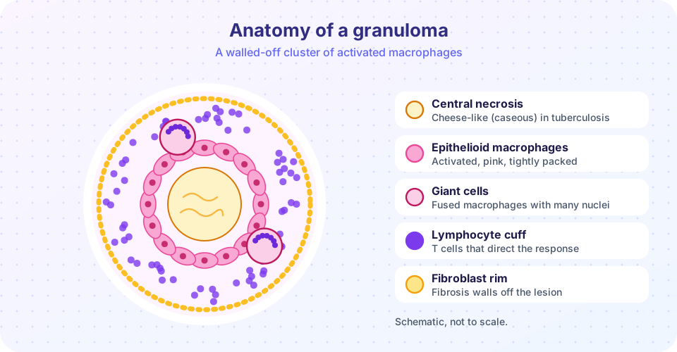
A granuloma is a pattern of chronic inflammation in which activated macrophages form a tight, nodular cluster. Under the microscope, the macrophages have pink, abundant cytoplasm and oval nuclei and are called epithelioid macrophages. They may fuse into multinucleated giant cells. In tuberculosis the nuclei of Langhans giant cells are arranged in a horseshoe around the rim. Around the cluster is a collar of lymphocytes, and older granulomas have a rim of fibroblasts and collagen.
There are two broad types:
- Immune granulomas are caused by persistent, poorly degradable particles that trigger a T-cell mediated immune reaction. Tuberculosis is the classic case.
- Foreign body granulomas form around inert material such as sutures, talc or splinters, without a major immune component. Macrophages and giant cells surround the object.
Tuberculosis as the model
When Mycobacterium tuberculosis is inhaled, alveolar macrophages ingest it. The bacterium resists killing by blocking the fusion of the phagosome with the lysosome. The infected macrophages present antigens and signal to T cells, which release interferon-gamma and activate the macrophages. The result is a granuloma that contains the infection. In many people the granuloma controls the bacteria for life; the bacteria may remain dormant inside it. The centre of a tuberculous granuloma undergoes caseous necrosis, giving a soft, cheese-like appearance (see the previous chapter). If immunity declines (HIV infection, drugs, malnutrition, aging), the infection can reactivate and spread. Robert Koch identified the tubercle bacillus in 1882.
Not all granulomas mean tuberculosis. Sarcoidosis (cause unknown) produces non-caseating granulomas, usually in the lungs and lymph nodes. Crohn disease, leprosy, some fungal infections and reactions to some drugs or to beryllium also form granulomas. The pathologist uses special stains and cultures to find the cause.
Consequences of chronic inflammation
- Fibrosis and loss of function. Scarring can stiffen or obstruct organs: cirrhosis of the liver after chronic hepatitis, narrowing of the intestine in Crohn disease, joint destruction in arthritis.
- Systemic effects. Fever, weight loss, fatigue, anemia of chronic disease (in which inflammatory signals raise hepcidin and trap iron), and raised CRP and ESR.
- Cancer risk. Chronic inflammation constantly stimulates cell division and produces damaging oxygen radicals. Chronic hepatitis B and C increase the risk of liver cancer, long-standing ulcerative colitis raises the risk of colon cancer, and Helicobacter pylori infection is linked with stomach cancer.
- Atherosclerosis. Seen as chronic inflammation of the artery wall, in which macrophages take up cholesterol and become foam cells.
Common misconceptions
- "A granuloma means tuberculosis." Many diseases form granulomas, and the cause needs further tests.
- "Chronic inflammation is just acute inflammation that lasted longer." The cells, mediators and tissue effects are different.
- "Inflammation in the body is always something to suppress." Chronic inflammation harms, but acute inflammation protects, and drugs that suppress it increase infection risks.
Try it
The first block is a toy model of the macrophage feedback loop. When the macrophage kills the microbe effectively, antigen is cleared and the inflammation resolves; when the microbe resists, the loop persists. The equations and numbers are invented to illustrate the idea.
Press Run (or Ctrl + Enter) to see the result here.
Program input · what you'd type while the program runs
Type the answers before you press Run, one per line. Each time the program reads a line (with input()), it gets the next line from this box.
The second block lists conditions with granulomas and the feature a pathologist looks for. Add your own rows as you meet new diseases.
Press Run (or Ctrl + Enter) to see the console output here.
No output.
Check your understanding
Quick check · 5 questions
of 5 answered You scored / 5 · perfect!
-
1. Which cells predominate in chronic inflammation?
Chronic inflammation is mononuclear: macrophages, lymphocytes and plasma cells. -
2. Which cytokine from T cells activates macrophages to kill microbes?
Th1 cells release interferon-gamma, which drives classical macrophage activation. -
3. What is a caseating granuloma typical of?
Tuberculous granulomas have a necrotic, cheese-like center. -
4. Which statement about M2 macrophages is correct?
Alternative activation favors repair; the two states form a spectrum. -
5. Chronic hepatitis B and C increase the risk of which cancer?
Constant inflammation and repair in the liver raise the risk of hepatocellular carcinoma.
Summary
- Chronic inflammation lasts weeks to years when the cause persists, with mononuclear cells, tissue destruction and attempted repair together.
- Persistent infection, toxic particles, autoimmunity and metabolic disease are common causes.
- Macrophages and T cells sustain each other in a feedback loop through interferon-gamma and interleukin-12.
- Granulomas, with epithelioid macrophages and giant cells, wall off persistent agents; tuberculosis forms caseating granulomas.
- Consequences include fibrosis, systemic effects and increased cancer risk.
Next: Healing and Repair
Track your progress
Create a free account to tick off lessons and pick up where you left off on any device.
of Pathology lessons completed