


















Lesson 33 of 46 · Gastrointestinal, Renal and Endocrine Pathology
Kidney disease: glomerular disease, failure, stones and tumors
Understand nephritic and nephrotic syndromes, acute and chronic kidney failure, pyelonephritis, kidney stones and kidney tumors through mechanism and pathology.
The kidneys receive about a fifth of the heart's output and filter the entire plasma volume many times a day. They remove waste (urea, creatinine), control water and salt, balance acid, and make hormones that regulate blood pressure and red cell production. Because the filter is a delicate mesh of tiny capillaries, it is vulnerable to the immune system, to high blood pressure and to diabetes. Because it depends on blood flow, it also fails when blood pressure drops.
A helpful way to organize kidney pathology is by where the problem sits. Before the kidney (prerenal): not enough blood arrives. In the kidney (intrinsic): the glomeruli, tubules, interstitium or vessels are damaged. After the kidney (postrenal): urine cannot drain. This lesson uses that scheme and then looks at specific diseases.
What you will learn
- The structure of the glomerular filter and how it fails
- The difference between nephritic and nephrotic syndromes
- The causes and course of acute kidney injury and chronic kidney disease
- How infection, stones and tumors affect the urinary tract
Key terms
Glomerular filtration rate (GFR): how much blood plasma the kidneys filter per minute, the best overall measure of kidney function (normal in young adults about 100 to 120 mL/min per 1.73 m²).
Proteinuria / hematuria: protein / blood in the urine.
Azotemia / uremia: rise of urea and creatinine in blood / the clinical syndrome that follows.
Oliguria: low urine output.
How the glomerulus works, and fails
Blood enters the glomerulus, a tuft of capillaries inside a cup called Bowman's capsule. The filter has three layers: the fenestrated capillary endothelium, the glomerular basement membrane (GBM), and the podocytes, cells whose foot processes interlock to leave narrow slits. The filter is size- and charge-selective: water and small molecules pass, while large proteins like albumin and blood cells stay in.
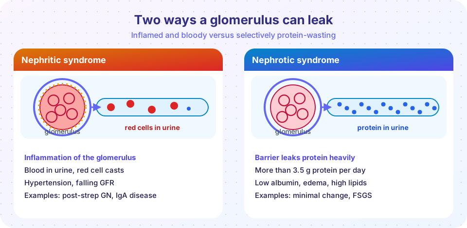
Disease can disturb the filter in two broad ways, giving two classic clinical pictures.
Nephritic syndrome
In the nephritic syndrome the glomerulus is inflamed. The signs are hematuria (red cells, often in casts, in dark "cola-colored" urine), a drop in GFR, hypertension from salt and water retention, and mild to moderate proteinuria. Examples:
- Post-streptococcal glomerulonephritis follows a streptococcal infection of the throat or skin, mostly in children. Immune complexes deposit under the podocytes (subepithelial humps), complement is consumed, and neutrophils swell the capillaries. Most children recover.
- IgA nephropathy is among the commonest glomerulonephritis in the world: IgA deposits in the mesangium and often cause visible hematuria a day or two after an upper respiratory infection.
- Lupus nephritis is kidney disease from immune complexes in systemic lupus erythematosus.
- Rapidly progressive glomerulonephritis (RPGN) is a severe form in which proliferating cells form crescents in Bowman's space, and kidney function can be lost in weeks. Causes include antibodies against the basement membrane (Goodpasture syndrome, which also bleeds into the lungs), ANCA-associated small-vessel vasculitis, and immune complexes.
Nephrotic syndrome
In the nephrotic syndrome the filter's selectivity fails and more than about 3.5 grams of protein are lost per day. The consequences follow from the loss: low blood albumin, edema from reduced oncotic pressure (often swollen eyelids and legs), high cholesterol (the liver makes more lipoproteins), and a tendency toward infections and blood clots (because antibodies and clotting inhibitors leak out). Major causes:
- Minimal change disease, the commonest cause in children. The glomeruli look normal under the light microscope; electron microscopy shows effacement of podocyte foot processes. It usually responds to corticosteroids.
- Focal segmental glomerulosclerosis (FSGS): scarring of parts of some glomeruli, from genetic variants, HIV, obesity or unknown cause.
- Membranous nephropathy: antibody deposits thicken the basement membrane, often with antibodies to the phospholipase A2 receptor on podocytes, in adults.
- Diabetic nephropathy: the leading cause of end-stage kidney disease. Long-standing high glucose thickens the basement membrane and expands the mesangium, producing Kimmelstiel-Wilson nodules and hyaline arteriolosclerosis.
Acute kidney injury
Acute kidney injury (AKI) is a rapid fall in kidney function over hours to days, causing a rise in creatinine and often a fall in urine output.
- Prerenal: reduced perfusion from dehydration, bleeding, heart failure or sepsis. The kidney tissue is intact, and function recovers when flow is restored.
- Intrinsic: the most common form is acute tubular necrosis (ATN), caused by ischemia or toxins (some antibiotics, contrast dye, heavy metals). Tubular cells die and slough off. ATN typically goes through an initiating phase, a maintenance phase with low urine output, and a recovery phase with a large urine volume while tubular cells regenerate. Other intrinsic causes are acute glomerulonephritis and acute interstitial nephritis, an allergic reaction to drugs.
- Postrenal: obstruction anywhere from the pelvis to the urethra, for example an enlarged prostate or stones, causes back-pressure and hydronephrosis.
Chronic kidney disease
Chronic kidney disease (CKD) is a lasting reduction in GFR (below 60 mL/min per 1.73 m² for three months or more) or evidence of kidney damage. The most common causes are diabetes and hypertension, followed by glomerulonephritis, polycystic disease and others. As nephrons are lost, remaining ones enlarge and filter more, which at first compensates but in the long run causes scarring, making the process self-perpetuating.
Complications reflect each lost kidney function: retention of urea and potassium, acidosis, anemia (low erythropoietin), hypertension, and mineral and bone disease (the kidney fails to activate vitamin D and to excrete phosphate, so calcium falls and the parathyroid glands overwork, causing secondary hyperparathyroidism). The end stage, with symptoms of uremia, is treated by dialysis or kidney transplantation.
Press Run (or Ctrl + Enter) to see the result here.
Program input · what you'd type while the program runs
Type the answers before you press Run, one per line. Each time the program reads a line (with input()), it gets the next line from this box.
The curve above shows something important: serum creatinine is a late and nonlinear marker. A large share of function can be lost before the number leaves the "normal" range, because a hyperbolic relation links the two. This is why doctors also look at urine protein and at trends.
Infection, stones and tumors
Urinary tract infection and pyelonephritis
Infection ascends from the urethra and bladder, usually caused by Escherichia coli, and is more common in women because of a shorter urethra. Acute pyelonephritis is infection of the kidney: patchy abscesses of neutrophils in the interstitium and tubules, with fever, flank pain and white cells in the urine, sometimes in casts. Reflux of urine up the ureter (vesicoureteral reflux) in childhood is a cause of chronic scarring.
Kidney stones
Most stones are calcium oxalate (about 80 percent). Others are struvite (magnesium ammonium phosphate, formed in infections with urease-producing bacteria such as Proteus), uric acid and cystine. Stones form when urine is supersaturated, with too little volume, too much of a stone-forming substance, or altered acidity. A stone lodged in the ureter causes severe, colicky pain, blood in urine and obstruction, which can lead to hydronephrosis.
Tumors
- Renal cell carcinoma is the main adult kidney cancer. The commonest type is clear cell carcinoma, with cells filled with lipid and glycogen that appear clear. Loss of the VHL gene is typical (inherited in von Hippel-Lindau syndrome). Smoking and obesity are risk factors. The classic signs, blood in urine, flank pain and a mass, are seen together in a minority. It can invade the renal vein and may make hormones, such as erythropoietin (a high red cell count) or PTHrP (high calcium).
- Urothelial carcinoma arises from the lining of the renal pelvis, ureter or bladder. Smoking and some industrial chemicals (aromatic amines) are major causes, and painless blood in the urine is the common presentation.
- Wilms tumor is the common kidney tumor of young children.
Common misconceptions
- "Blood in the urine means kidney disease." It can come from anywhere in the urinary tract, including stones, infection and tumors.
- "A normal creatinine means normal kidneys." Creatinine is insensitive early on; protein in urine can reveal damage before creatinine rises.
- "Nephrotic syndrome and nephritic syndrome are the same." One is mainly protein loss, the other mainly inflammation with blood and hypertension.
- "Kidney stones come from drinking milk." Calcium intake in food does not appear to be the main driver; low fluid intake is a bigger factor.
Try it
Press Run (or Ctrl + Enter) to see the console output here.
No output.
Check your understanding
Quick check · 5 questions
of 5 answered You scored / 5 · perfect!
-
1. Which feature is typical of nephrotic syndrome?
Heavy proteinuria, hypoalbuminemia, edema and hyperlipidemia define the nephrotic syndrome. -
2. The commonest cause of nephrotic syndrome in children is
Minimal change disease shows normal glomeruli by light microscopy and foot process effacement by EM. -
3. In acute tubular necrosis the maintenance phase typically shows
Oliguria dominates the maintenance phase, followed by a polyuric recovery phase. -
4. Which two diseases are the leading causes of chronic kidney disease?
Diabetic and hypertensive kidney disease account for most CKD. -
5. The most common kidney stone type is
About 80 percent of stones contain calcium, mostly as oxalate.
Summary
- Kidney disease can be prerenal, intrinsic or postrenal; the glomerular filter is a three-layer barrier.
- Nephritic syndrome: inflamed glomeruli, hematuria, hypertension. Nephrotic syndrome: heavy proteinuria, edema, low albumin.
- AKI often means ATN; CKD is most often from diabetes and hypertension, with anemia, acidosis and bone disease.
- Pyelonephritis, stones and kidney tumors each have recognizable causes and presentations.
Next: Endocrine disease
Track your progress
Create a free account to tick off lessons and pick up where you left off on any device.
of Pathology lessons completed