


















Lesson 12 of 46 · Inflammation and Repair
Acute Inflammation: Vessels, White Cells and Mediators
Learn the vascular and cellular steps of acute inflammation: vasodilation, leaky vessels, leukocyte recruitment, phagocytosis, mediators and the common patterns.
Acute inflammation is the fast, front-line response to injury. It starts within minutes, builds over hours, and is usually over within days. Two things happen in sequence. First, blood vessels change so that plasma proteins and white cells can reach the tissue. Second, white cells leave the vessels, travel to the problem, and destroy or remove it. Knowing these steps lets you predict what an inflamed tissue will look like, why different drugs help, and what goes wrong in diseases where a step is missing.
What you will learn
- The vascular changes of acute inflammation and why they cause swelling.
- How exudate and transudate differ.
- The five steps by which white cells leave the blood.
- How phagocytes kill microbes, and what happens when they cannot.
- The main chemical mediators and common morphological patterns.
Key terms
Exudate: fluid with high protein content and cells that leaks from inflamed vessels.
Transudate: protein-poor fluid that leaks because of pressure imbalance, without vessel injury.
Chemotaxis: movement of cells along a chemical gradient.
Opsonin: a coating (antibody or complement) that marks a microbe for phagocytosis.
Respiratory burst: rapid production of reactive oxygen species by phagocytes.
Part 1: vascular changes
Vasodilation
An injury is followed by a brief narrowing of arterioles, then a much longer widening, driven largely by histamine and nitric oxide. The widened arterioles open up the capillary beds, so blood flow to the area rises. A simple physical fact explains how large the effect can be: flow through a tube of radius r is proportional to r⁴ (the Poiseuille relationship), so a small increase in radius gives a large increase in flow. This is the source of redness and heat.
Increased vascular permeability
The most important change is that the smallest vessels, especially venules, become leaky. Several mechanisms exist:
- Endothelial contraction. Histamine, bradykinin and leukotrienes cause endothelial cells to contract and open gaps between them. This is rapid (minutes) and short-lived.
- Direct endothelial injury. Burns, some toxins and severe trauma kill endothelial cells, producing leakage that begins immediately and lasts until the vessels are repaired or thrombosed.
- Leukocyte-mediated injury. White cells that have adhered to the vessel wall release enzymes and oxygen radicals that damage it.
- Increased transcytosis. Some mediators (such as vascular endothelial growth factor) increase transport of fluid across the endothelial cell in vesicles.
The result is exudate, protein-rich fluid that moves into the tissue because the protein raises the tissue's osmotic pull and the lost proteins no longer hold fluid in the vessel. The tissue swells (the edema of "tumor"). Compare this with a transudate, such as the fluid of heart failure edema, which is protein-poor because the vessel wall is intact and the fluid is pushed out only by raised pressure. Telling exudate from transudate by laboratory analysis can show whether the cause is inflammation or a pressure disorder.
Slowing of flow
As fluid leaves, the remaining blood becomes more concentrated and more viscous, and flow slows (stasis). This helps white cells move from the center of the stream to the vessel wall.
Part 2: leukocyte recruitment
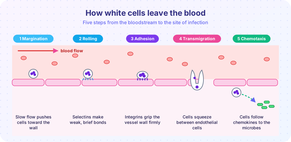
To reach tissue, white cells must leave the circulation. Five steps do it.
- Margination. In normal flow, cells travel in the center of the vessel. When flow slows, white cells drift toward the wall.
- Rolling. Selectins on the endothelium (P-selectin and E-selectin, switched on by histamine and cytokines such as TNF and interleukin-1) bind sugar structures on the leukocyte. The bonds are weak and brief, so the cell rolls along the wall.
- Firm adhesion. Chemokines presented on the endothelial surface activate integrins on the leukocyte (such as LFA-1 and Mac-1), switching them to a high-affinity form. They bind ICAM-1, which cytokines have increased on the endothelium. The cell stops.
- Transmigration (diapedesis). The cell squeezes between endothelial cells, helped by molecules such as PECAM-1 (CD31), and crosses the basement membrane by secreting enzymes.
- Chemotaxis. Once outside, leukocytes follow a gradient of chemoattractants (bacterial products, the complement fragment C5a, leukotriene B4 and chemokines such as interleukin-8) toward the source.
In the first 6 to 24 hours, neutrophils dominate. After 24 to 48 hours, monocytes arrive and mature into macrophages. The switch occurs partly because neutrophils live only a day or two and partly because the chemokines produced change with time. An exception: in viral infections, lymphocytes may predominate first, and in allergic reactions and parasitic infections, eosinophils.
Genetic diseases show how essential the steps are. In leukocyte adhesion deficiency type 1, a defect in the integrin subunit CD18 prevents firm adhesion. Patients suffer repeated bacterial infections, produce no pus, and their blood is full of neutrophils that cannot leave it.
Part 3: phagocytosis and killing
Once at the site, neutrophils and macrophages must kill the microbe.
- Recognition and attachment. Phagocytes bind microbes directly through receptors, or more efficiently when the microbe is coated with opsonins: antibodies (IgG) and the complement fragment C3b.
- Engulfment. The cell membrane wraps around the particle, forming a vesicle called a phagosome, which fuses with lysosomes to form a phagolysosome.
- Killing. The main weapon is the respiratory burst: the enzyme NADPH oxidase converts oxygen to superoxide, which becomes hydrogen peroxide. The enzyme myeloperoxidase (MPO), stored in neutrophil granules, uses hydrogen peroxide and chloride to produce hypochlorous acid, essentially bleach, which kills bacteria. Lysosomal enzymes (proteases, lysozyme) and nitric oxide help.
In chronic granulomatous disease NADPH oxidase is missing, so killing is impaired; patients suffer severe infections with certain bacteria and fungi and form granulomas (see the next lesson). Neutrophils can also cast out web-like structures called neutrophil extracellular traps (NETs), made of DNA and antimicrobial proteins, which trap microbes but may also injure the host.
Phagocytes sometimes damage innocent tissue because their weapons leak into the surroundings. This is how inflammation causes collateral damage.
Part 4: chemical mediators
Mediators explain most of the previous steps. This table lists the main ones.
| Mediator | Source | Main actions |
|---|---|---|
| Histamine | Mast cells | Vasodilation, increased permeability (early) |
| Prostaglandins | Many cells (via COX enzymes) | Vasodilation, pain, fever |
| Leukotrienes | Leukocytes, mast cells | Increased permeability, chemotaxis; some cause bronchoconstriction |
| TNF and IL-1 | Macrophages | Endothelial activation, fever, acute-phase response |
| Chemokines | Many cells | Chemotaxis, leukocyte activation |
| Complement (C3a, C5a, C3b) | Plasma proteins | Increased permeability, chemotaxis, opsonization |
| Bradykinin | Kinin system | Pain, increased permeability |
Common drugs act on these pathways. Aspirin and other non-steroidal anti-inflammatory drugs (NSAIDs) block the cyclooxygenase enzymes that make prostaglandins, relieving pain and fever. Corticosteroids have broad effects, including reducing production of many mediators. Antihistamines block the actions of histamine on its receptors.
Part 5: patterns you will see
- Serous inflammation: thin fluid, as in a skin blister.
- Fibrinous inflammation: large leaks of fibrinogen, which forms fibrin sheets on surfaces, as in pericarditis, where the heart's lining looks "bread and butter" rough.
- Suppurative (purulent) inflammation: pus, made of dead neutrophils, debris and fluid. A localized collection is an abscess.
- Ulcer: local loss of a surface lining, with inflammation of the underlying tissue, as in a stomach ulcer.
Common misconceptions
- "Pus means the infection is getting worse." Pus is the product of a working neutrophil response. A large collection may need drainage, but its presence shows the body is responding.
- "Swelling is mainly extra blood." Most swelling is fluid and protein leaking from small vessels.
- "Anti-inflammatory drugs are only for pain." They reduce several mediator-driven features, including fever and swelling.
Try it
The first block illustrates the vessel-radius relationship that makes small changes in diameter matter. The values are relative; it is a simplified physical demonstration.
Press Run (or Ctrl + Enter) to see the result here.
Program input · what you'd type while the program runs
Type the answers before you press Run, one per line. Each time the program reads a line (with input()), it gets the next line from this box.
The second block is a rule-of-thumb sorter for exudate and transudate. The cut-offs are simplified textbook values, and real fluid analysis uses more criteria.
Press Run (or Ctrl + Enter) to see the console output here.
No output.
Check your understanding
Quick check · 6 questions
of 6 answered You scored / 6 · perfect!
-
1. Which molecules mediate the rolling step of leukocyte recruitment?
Selectins make weak, brief bonds that make leukocytes roll. Integrins mediate firm adhesion. -
2. A fluid sample has high protein and many cells. This suggests:
Exudate is protein-rich and often cellular because vessels have become leaky. -
3. What is the main killing mechanism of neutrophils in a phagolysosome?
NADPH oxidase and myeloperoxidase produce reactive oxygen species and hypochlorous acid. -
4. Which drug class mainly blocks prostaglandin production?
NSAIDs inhibit cyclooxygenase enzymes. -
5. In leukocyte adhesion deficiency type 1, which problem occurs?
A defect in CD18 integrins blocks firm adhesion and transmigration. -
6. If a vessel's radius increases by 20 percent at constant pressure, the flow increases roughly by:
Flow scales with r to the fourth power, and 1.2 to the fourth is about 2.07.
Summary
- Acute inflammation begins with vasodilation and increased permeability, producing exudate and swelling.
- Leukocytes leave the blood by margination, rolling (selectins), adhesion (integrins), transmigration and chemotaxis.
- Neutrophils come first, followed by monocytes and macrophages.
- Phagocytes recognize, engulf and kill microbes using the respiratory burst, MPO and lysosomal enzymes.
- Histamine, prostaglandins, cytokines, chemokines, complement and kinins coordinate the response.
- Patterns include serous, fibrinous and suppurative inflammation, abscesses and ulcers.
Track your progress
Create a free account to tick off lessons and pick up where you left off on any device.
of Pathology lessons completed