


















Lesson 9 of 46 · Cell Injury and Death
Necrosis and Apoptosis: How Cells Die
Compare necrosis and apoptosis, learn the five patterns of necrosis, the caspase pathways of programmed cell death, and when too little or too much death causes disease.
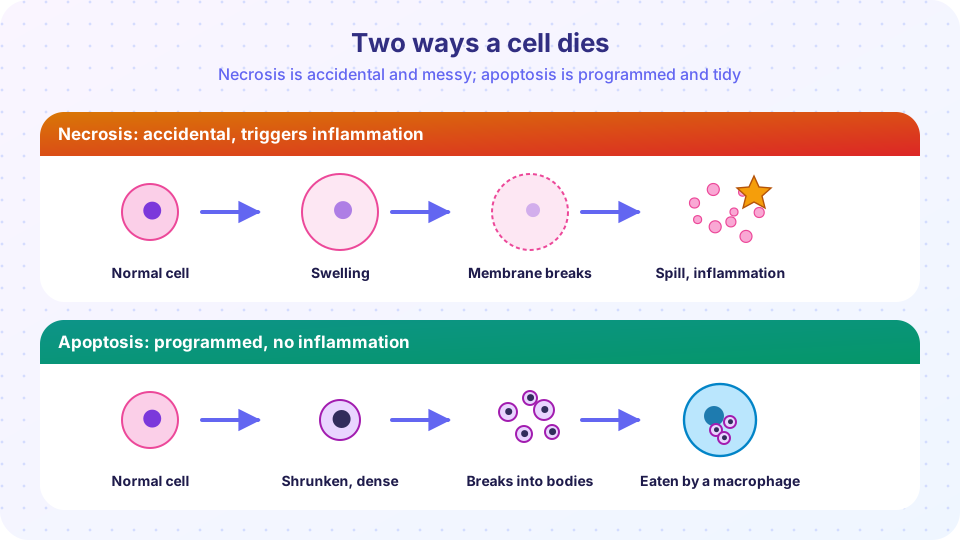
Cells die in two broad styles. Some are killed by an injury they cannot survive: their membranes burst, their contents spill, and the neighboring tissue reacts with inflammation. This is necrosis. Others carry out a self-destruction program that dismantles them quietly and neatly, without alarming the immune system. This is apoptosis, often called programmed cell death. The difference matters in medicine because it decides whether tissue dies noisily (with inflammation, pain and scar) or silently, and because many diseases, including cancer and autoimmune disease, involve a breakdown in the control of cell death.
What you will learn
- The nuclear and cytoplasmic changes of necrosis, and the five main patterns.
- What apoptosis is for, in health and disease.
- The two caspase pathways that run apoptosis.
- A short guide to other forms of regulated cell death.
Key terms
Necrosis: death of cells caused by injury, with membrane rupture and inflammation.
Apoptosis: energy-dependent, regulated cell suicide in which the membrane stays intact.
Caspases: protein-cutting enzymes that carry out apoptosis.
Apoptotic bodies: membrane-bound fragments of a dying cell, taken up by phagocytes.
Pyknosis, karyorrhexis, karyolysis: shrinkage, fragmentation and fading of the nucleus.
Necrosis
Necrosis is what you see when the injury is too severe for the cell to survive, such as a long interruption of blood supply, a toxin or a burn. Two processes run together: enzymatic digestion of the cell (by its own lysosomal enzymes, or by those of white cells that arrive) and denaturation of proteins. The plasma membrane loses integrity, and cell contents leak out. The spilled molecules are recognized as danger signals and recruit inflammatory cells.
Under the microscope the cytoplasm looks more intensely pink (eosinophilic) because it has lost its RNA and the proteins are denatured. The nucleus shows one of three changes:
- Pyknosis: the nucleus shrinks and becomes a dense, dark ball.
- Karyorrhexis: the pyknotic nucleus breaks into pieces.
- Karyolysis: the nucleus fades as DNA is digested.
Patterns of necrosis
Pathologists name necrosis by its gross and microscopic pattern, which helps identify the cause.
| Pattern | What it looks like | Typical setting |
|---|---|---|
| Coagulative | Tissue stays firm; outlines of cells survive for days as "ghosts" | Ischemic death of solid organs (heart, kidney, spleen) |
| Liquefactive | Tissue dissolves into a liquid, often pus | Bacterial infections (abscesses) and brain infarcts |
| Caseous | Soft, crumbly, white or cheese-like, structureless | Tuberculosis |
| Fat necrosis | Chalky white areas where fat has been digested | Acute pancreatitis; trauma to fat |
| Fibrinoid | Bright pink, fibrin-like material in vessel walls | Immune reactions in blood vessels, severe hypertension |
Why is the brain different? Coagulative necrosis is the typical outcome of ischemia in most organs because the same injury that kills the cells also denatures the enzymes that would digest them. Brain tissue contains a lot of fat and is rich in digestive enzymes and microglia, so an infarct of the brain liquefies. In acute pancreatitis, pancreatic enzymes called lipases leak into surrounding fat and split triglycerides into fatty acids that bind calcium, forming white chalky soaps (saponification).
Gangrene is a clinical term rather than a pattern. It describes necrosis of a limb or organ, usually from loss of blood supply. Dry gangrene is coagulative necrosis; wet gangrene involves bacterial superinfection and liquefactive features.
Apoptosis
Apoptosis (from a Greek word for leaves falling) is a controlled process, controlled by genes and requiring energy. Its hallmarks:
- the cell shrinks and its cytoplasm becomes dense,
- the chromatin condenses and the nucleus breaks into fragments,
- the plasma membrane forms blebs and the cell separates into apoptotic bodies that retain intact membranes,
- the bodies display "eat-me" signals (such as phosphatidylserine on the outer surface), so phagocytes remove them quickly,
- there is no leakage of contents and therefore no inflammation.
Apoptosis in health
In a healthy body, huge numbers of cells die by apoptosis every day. Examples:
- Development. Webbing between the fingers of a fetus disappears through apoptosis. The nervous system produces more neurons than are needed, and the surplus dies.
- Tissue turnover. The lining of the gut is shed and replaced every few days.
- Hormone withdrawal. The uterine lining falls apart when progesterone falls; the breast regresses after lactation.
- Immune system. Lymphocytes that react against the body's own tissues are removed, and when an infection ends, excess immune cells are deleted.
Apoptosis in disease
Apoptosis also fires when cells are damaged beyond repair: severe DNA damage activates the protein p53, which can trigger apoptosis rather than allow a possibly cancerous cell to survive. Misfolded proteins (ER stress), some viral infections (a viral hepatitis shows scattered shrunken apoptotic liver cells called Councilman bodies) and duct obstruction (which causes atrophy of the organ behind the block) all involve it.
How apoptosis works
Apoptosis is run by a family of enzymes called caspases. Caspases exist as inactive precursors that are activated by cutting. There are two main routes in, and both end at the same executioners.
The intrinsic (mitochondrial) pathway
Stresses such as DNA damage or lack of growth factors tip the balance of the BCL-2 family of proteins. Anti-apoptotic members (BCL-2, BCL-XL) keep the mitochondrial membrane sealed. Pro-apoptotic members (BAX, BAK) form pores. When pores open, cytochrome c leaks from mitochondria into the cytoplasm, joins a protein called APAF-1 to form the apoptosome, and activates caspase-9.
The extrinsic (death receptor) pathway
Death receptors on the cell surface (Fas, and the TNF receptor) are triggered by ligands from other cells. For example, cytotoxic T cells carry Fas ligand. Receptor binding assembles an adaptor protein (FADD) that activates caspase-8.
The execution phase
Both routes activate the executioner caspases (3, 6 and 7), which cut hundreds of cell proteins and activate DNases that cleave DNA between nucleosomes into fragments, in multiples of about 180 base pairs. On a gel this gives a characteristic "ladder" pattern, which is used as a laboratory test for apoptosis.
When the balance goes wrong
Tissue size is the net result of cell birth and cell death. Too little apoptosis lets abnormal cells survive: cancers often have a defect in the process (in follicular lymphoma, a translocation overexpresses BCL-2), and failure to remove self-reactive lymphocytes contributes to autoimmune disease. Too much apoptosis causes loss of tissue: the death of neurons in neurodegenerative diseases, loss of CD4 T cells in HIV infection, and part of the cell loss after ischemia.
Other forms of cell death
Cell death is more varied than the old necrosis versus apoptosis split. Three more regulated forms are worth knowing.
- Necroptosis: a regulated form of necrosis that can occur when caspase-8 is blocked. It requires proteins named RIPK1, RIPK3 and MLKL, which punch holes in the membrane. It causes inflammation.
- Pyroptosis: death driven by inflammasomes and caspase-1, in which gasdermin proteins form pores. It releases inflammatory signals and is a defense against some infections.
- Ferroptosis: death caused by iron-dependent oxidation of membrane lipids.
Autophagy, mentioned in the atrophy lesson, is mostly a survival mechanism, though prolonged autophagy can lead to cell death.
Common misconceptions
- "Apoptosis is just a kind of disease." It is a normal and essential process; billions of cells die by apoptosis in a healthy adult every day.
- "Necrosis and apoptosis are exclusive." Many injuries cause both, and a dying cell can switch between forms depending on how severe the stress is and how much ATP is left.
- "Necrosis is the same as gangrene." Gangrene is a clinical description of dead tissue; necrosis is the process, with several patterns.
Try it
The first block simulates how the balance between cell birth and apoptosis sets tissue size. The rates are invented for demonstration.
Press Run (or Ctrl + Enter) to see the result here.
Program input · what you'd type while the program runs
Type the answers before you press Run, one per line. Each time the program reads a line (with input()), it gets the next line from this box.
The second block matches settings to patterns of necrosis, as in the table.
Press Run (or Ctrl + Enter) to see the console output here.
No output.
Check your understanding
Quick check · 6 questions
of 6 answered You scored / 6 · perfect!
-
1. Which feature distinguishes apoptosis from necrosis?
In apoptosis the cell shrinks into membrane-bound fragments that phagocytes remove, so there is no inflammation. -
2. A pathologist sees a pale, soft, crumbly, cheese-like area in a lung. Which pattern is this?
Caseous necrosis is typical of tuberculosis. -
3. What does a pyknotic nucleus look like?
Pyknosis is shrinkage and darkening; karyorrhexis is fragmentation; karyolysis is fading. -
4. Which molecule released from mitochondria starts the apoptosome in the intrinsic pathway?
Cytochrome c binds APAF-1 and activates caspase-9. -
5. Overexpression of BCL-2 in a lymphoma tends to cause what?
BCL-2 blocks apoptosis, so cells that should die persist. -
6. Why does an infarct of the brain typically undergo liquefactive necrosis?
Brain tissue is rich in lipid and enzymes, so dead tissue dissolves rather than staying firm.
Summary
- Necrosis follows irreversible injury, spills cell contents and causes inflammation; its nuclear changes are pyknosis, karyorrhexis and karyolysis.
- Patterns: coagulative, liquefactive, caseous, fat and fibrinoid, each linked to typical settings.
- Apoptosis is programmed, energy-dependent and silent; it shapes development, removes damaged cells and regulates the immune system.
- Caspases carry out apoptosis through the intrinsic (BCL-2 and cytochrome c) and extrinsic (death receptor) pathways.
- Too little apoptosis favors cancer and autoimmunity; too much causes tissue loss. Necroptosis, pyroptosis and ferroptosis are other regulated types.
Next: Chapter 2 Review
Track your progress
Create a free account to tick off lessons and pick up where you left off on any device.
of Pathology lessons completed