


















Lesson 22 of 46 · Neoplasia
Names and growth: benign, malignant and metastasis
Learn how tumors are named, how benign and malignant growth differ, what dysplasia and anaplasia mean, and how metastasis happens step by step.
Pathology has a naming system for tumors that, once you see the logic, lets you decode a diagnosis from the word alone. The system rests on two questions: what kind of cell does the tumor come from, and does it behave in a benign or malignant way? Answering them also tells you a lot about what will happen next, because a tumor that grows in place is a very different problem from one that invades and travels.
This lesson builds the vocabulary, the microscopic signs of malignancy, and the step-by-step process by which a cancer spreads. Along the way you will meet the famous exceptions to the rules, because pathology is full of them.
What you will learn
- How to read a tumor name: tissue of origin plus behavior
- The microscopic differences between benign and malignant growth
- What differentiation, dysplasia and anaplasia mean
- The routes and steps of metastasis, and why most cells fail
Key terms
Differentiation: how closely tumor cells resemble the normal cells they come from.
Anaplasia: lack of differentiation, a sign of malignancy.
Dysplasia: disordered, premalignant epithelial growth that has not invaded.
Invasion: growth into and destruction of neighboring tissue.
Metastasis: a tumor that arises from cells that traveled from the primary tumor.
Reading a tumor's name
A tumor has two parts to its name: a root from its tissue of origin, and an ending that signals its behavior.
- Benign tumors usually end in -oma: a benign tumor of fat is a lipoma, of smooth muscle a leiomyoma, of glandular epithelium an adenoma, and of bone an osteoma.
- Malignant tumors of epithelium are carcinomas. Those of glandular epithelium are adenocarcinomas, and those of squamous epithelium are squamous cell carcinomas.
- Malignant tumors of mesenchyme (connective tissue, muscle, fat, bone) are sarcomas: liposarcoma, leiomyosarcoma, osteosarcoma, angiosarcoma.
- Blood-forming cells give leukemias (in the blood and marrow) and lymphomas (solid masses of lymphoid tissue).
The tissue categories matter because the three big families of cancer behave differently. Carcinomas are the most common cancers in adults and tend to spread first through lymphatic vessels. Sarcomas are rarer and tend to spread through the blood. Roots also tell you about structure: a papilloma is a finger-like projection, a polyp projects into a lumen, and a cystadenoma is a glandular tumor with cysts.
The traps
Naming has exceptions you must memorize, because the word and the behavior do not always agree.
- Melanoma, lymphoma, mesothelioma and seminoma all end in -oma but are malignant.
- A teratoma arises from germ cells and contains tissues from more than one germ layer (hair, teeth and so on); mature teratomas of the ovary are usually benign, but immature ones can be malignant.
- A hamartoma is a disorganized mass of tissue native to the site (a lung "coin lesion" with cartilage is a classic). A choristoma is normal tissue in the wrong place. Neither is a true neoplasm in the usual sense.
Benign versus malignant: how pathologists tell
No single feature decides the question. Pathologists weigh several.
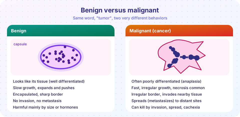
Differentiation and anaplasia. In a well-differentiated tumor the cells look and act like the tissue they come from: a benign adenoma may form neat glands. As tumors become more malignant, cells lose those features. A very poorly differentiated or anaplastic tumor may be so unlike its parent tissue that special stains are needed to find its origin. The hallmarks of anaplasia are:
- Pleomorphism: variation in cell and nuclear size and shape.
- A high nuclear-to-cytoplasmic ratio, because the nucleus is oversized (normally about 1:4 to 1:6).
- Hyperchromasia: dark nuclei with excess DNA.
- Abnormal mitoses, including multipolar spindles, and often many mitotic figures.
- Loss of polarity: cells grow in disorderly sheets rather than layers.
- Tumor giant cells and, in fast-growing tumors, areas of necrosis.
Rate of growth. Benign tumors tend to grow slowly, malignant tumors faster, but there are overlaps. A leiomyoma (fibroid) of the uterus can grow quickly during pregnancy because of hormones, while some cancers grow slowly for years. Rapid growth usually outpaces blood supply, which causes central necrosis.
Local invasion. A benign tumor expands and pushes neighbors aside, often forming a fibrous capsule. This is why many can be shelled out surgically. A malignant tumor infiltrates: it penetrates blood vessels, nerves and neighboring tissues, with no clean plane, which is why surgeons take a margin of normal tissue. The word "cancer" comes from the Greek and Latin for crab, supposedly because of the claw-like limbs of invasive growths.
Metastasis. Spread to a site not connected to the primary tumor is the most reliable sign of malignancy. There are rare exceptions for tumors that do not spread but are locally aggressive, such as basal cell carcinoma of the skin and most gliomas of the brain, which usually do not metastasize outside the nervous system.
A spectrum, not a switch
Many epithelial cancers do not appear suddenly. They are preceded by dysplasia, a disordered epithelium with abnormal cell size and shape, enlarged dark nuclei and lost layering. Mild dysplasia can regress if the cause (for example, an HPV infection or chronic irritation) is removed. When dysplastic cells fill the whole thickness of the epithelium but the basement membrane is still intact, the lesion is called carcinoma in situ. In situ is Latin for "in place." It is not yet invasive, which is why finding it through screening (such as cervical cytology) allows treatment before spread.
Two related changes are not neoplasia but are often confused with it. Hyperplasia is an increase in the number of cells due to a stimulus, such as thickening of the uterine lining under estrogen, and it usually reverses when the stimulus ends. Metaplasia is replacement of one mature cell type with another that is better suited to a stress, such as squamous metaplasia in the airways of smokers or the intestinal-type lining of Barrett esophagus after chronic acid reflux. Metaplasia is reversible in principle, but a metaplastic tissue exposed to a persistent insult can become a seedbed for dysplasia.
The route out: how metastasis works
Metastasis is a sequence of steps, often called the invasion-metastasis cascade, and each step is a filter that most cells fail.
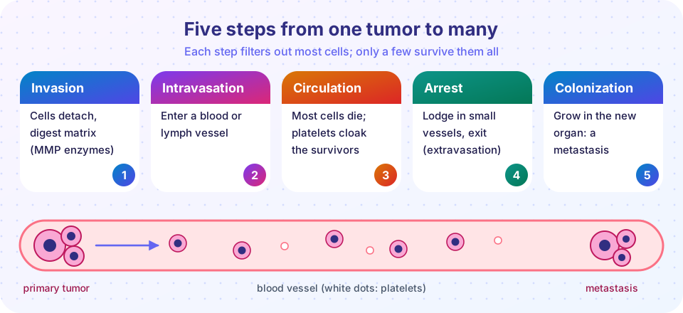
- Invasion of the matrix. Cells lose their adhesion to each other (a loss of E-cadherin is a classic change), attach to the extracellular matrix, and release matrix metalloproteinases to chew through it. They move.
- Intravasation. Cells enter the lumen of a lymphatic or blood vessel.
- Survival in circulation. Most cells die from shear forces and attack by immune cells. Tumor cells that clump with platelets are partly shielded.
- Arrest and extravasation. Cells lodge in small vessels, often at sites that match a particular tumor, and exit into the tissue.
- Colonization. At the new site cells must find a hospitable setting. Most that arrive never form a colony, and some stay dormant for years, which explains late recurrences.
Three routes
- Lymphatic spread is the usual first route for carcinomas. Cancer cells travel to regional lymph nodes first, which is why surgeons examine the sentinel node, the first node draining a tumor.
- Hematogenous (blood) spread is typical for sarcomas, but also for some carcinomas such as renal cell carcinoma, which can invade the renal vein, and hepatocellular carcinoma. Because veins drain to the liver or lungs first, these two organs are the most common sites for metastases. Cancers of the colon drain via the portal vein to the liver; many others go to the lungs. Bone is a frequent destination for prostate and breast cancers.
- Transcoelomic (seeding) spread occurs when cells cross a body cavity, such as ovarian cancer spreading across the peritoneum.
Why do cells go to particular organs? Two ideas, both from different centuries, combine. In 1889 Stephen Paget described the seed and soil hypothesis: certain cells (seeds) grow only in organs whose environment (soil) suits them. James Ewing, in the 1920s, argued for mechanics: blood flow and anatomy decide where cells get stuck. Modern work shows both matter, with signaling molecules guiding cells (chemokines, for example) to particular organs.
Common misconceptions
- "Benign means harmless." A benign tumor can still cause harm by pressing on organs (a meningioma in a closed skull), blocking a duct, or secreting hormones (an insulin-producing adenoma causes dangerous low blood sugar).
- "A tumor that spreads is a new cancer." A liver metastasis from colon cancer is still colon cancer; it is treated and named as such ("metastatic colon cancer").
- "All cancers form a lump." Leukemias do not, and some cancers spread through tissue (diffuse gastric cancer) without a distinct mass.
Try it
These two demonstrations are teaching models. They are not data from real patients.
Press Run (or Ctrl + Enter) to see the result here.
Program input · what you'd type while the program runs
Type the answers before you press Run, one per line. Each time the program reads a line (with input()), it gets the next line from this box.
Check your understanding
Quick check · 5 questions
of 5 answered You scored / 5 · perfect!
-
1. A malignant tumor of smooth muscle is called a
Mesenchymal malignant tumors end in -sarcoma; leiomyoma is the benign version. -
2. Which of these is malignant despite ending in -oma?
Melanoma, lymphoma, mesothelioma and seminoma are malignant despite their endings. -
3. Carcinoma in situ means
In situ means in place: the cells have not breached the basement membrane. -
4. Which finding most reliably marks a tumor as malignant?
Spread to a disconnected site is the most reliable sign; the other features overlap between benign and malignant. -
5. Carcinomas most often spread first by which route?
Carcinomas typically reach regional lymph nodes first; sarcomas tend to use the blood.
Summary
- Tumor names combine tissue of origin with behavior: -oma for benign, carcinoma for malignant epithelium, sarcoma for malignant mesenchyme.
- Beware of exceptions: melanoma, lymphoma, mesothelioma and seminoma are malignant.
- Anaplasia means pleomorphism, large dark nuclei, abnormal mitoses and loss of polarity.
- Dysplasia and carcinoma in situ come before invasion.
- Metastasis is a multistep cascade through lymphatic, blood or cavity routes, and most cells fail at each step.
Next: Carcinogenesis
Track your progress
Create a free account to tick off lessons and pick up where you left off on any device.
of Pathology lessons completed