


















Lesson 37 of 46 · Infectious and Neurological Disease
Host response to infection: granulomas, tuberculosis and sepsis
Learn the tissue patterns of infection, how tuberculosis forms caseating granulomas, how viruses injure cells, and how sepsis and septic shock develop.
An infection is not a one-sided attack. The microbe damages tissue directly, using toxins, enzymes and by killing the cells it lives in. The host damages tissue in turn, by flooding it with neutrophils, by walling off an invader with scar, or by releasing inflammatory mediators that leak into the whole circulation. Good pathology keeps both sides in view, because the pattern of tissue injury points to the microbe and the cost of the response explains why some infections are so dangerous.
This lesson goes through the common inflammatory patterns, then looks at the infection that most shaped modern thinking about granulomas, tuberculosis, and finally at the point where an infection stops being local: sepsis.
What you will learn
- The main tissue patterns of infection and what each suggests
- How the immune system forms a granuloma and why it sometimes fails
- The stages of tuberculosis, from primary infection to reactivation
- How viruses injure cells
- The cascade of events in sepsis, septic shock and disseminated intravascular coagulation
Key terms
Abscess: a localized collection of pus walled off by tissue.
Caseous necrosis: cheese-like, pale tissue death typical of tuberculosis.
Cytopathic effect: visible changes in cells caused by a virus.
Endotoxin: lipopolysaccharide in the outer wall of gram-negative bacteria that triggers strong immune responses.
Disseminated intravascular coagulation (DIC): widespread clotting in small vessels that uses up clotting factors and causes bleeding.
Patterns of tissue response
Think of the pattern as a clue.
- Suppurative (pyogenic) inflammation. Pyogenic bacteria such as Staphylococcus aureus and streptococci attract huge numbers of neutrophils. Neutrophil enzymes dissolve tissue (liquefactive necrosis), forming pus. A localized collection is an abscess. The same bacteria in the lung cause bronchopneumonia, and in the meninges they cause bacterial meningitis.
- Mononuclear and viral patterns. Viruses live inside cells, so the immune response is largely carried out by lymphocytes. Tissue shows lymphocytes cuffing small vessels, and cells that are dead, swollen, or marked by a virus: inclusion bodies (herpes simplex produces "ground glass" nuclei with eosinophilic inclusions; cytomegalovirus produces cells with an "owl's eye" nucleus; rabies produces Negri bodies in neurons), multinucleated giant cells from fused cells (herpes viruses, measles, respiratory syncytial virus), or koilocytes, which are cells with halos around shrunken nuclei in HPV infection. Some viruses stimulate cell growth (warts, molluscum).
- Granulomatous inflammation. Described below.
- Necrotizing infections. Clostridium perfringens releases toxins that destroy muscle and produce gas (gas gangrene); severe soft tissue infections spread along fascial planes (necrotizing fasciitis).
- Eosinophilic and chronic patterns. Parasites, especially worms, provoke eosinophils and IgE. Long-lasting infections end in scar and organ distortion, as in chronic hepatitis.
Granulomas
A granuloma is a response to an agent that macrophages cannot destroy: a bacterium protected by its wall, a fungus, a foreign body, or an unknown trigger. T helper cells recognize the antigen and secrete interferon-gamma, which turns macrophages into powerful killers and also transforms them into epithelioid cells, so named because their pink, tightly packed appearance resembles epithelium. Epithelioid cells may fuse into multinucleated giant cells, including the Langhans giant cell, whose nuclei line up in a horseshoe at the edge. The whole ball is ringed by lymphocytes and fibroblasts.
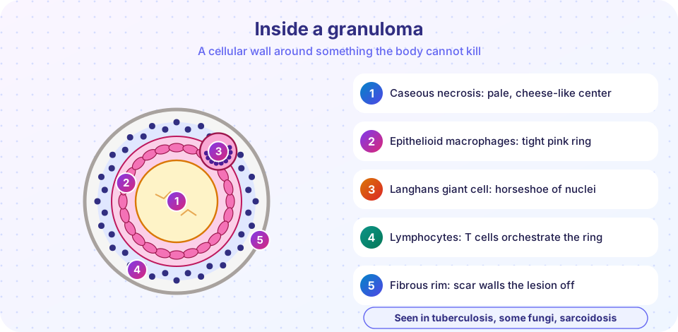
Granulomas are caseating when the center dies into pale, soft, cheese-like debris, which is typical of tuberculosis. They are non-caseating in sarcoidosis, Crohn disease and some other settings. Other causes include fungal infections (histoplasmosis), leprosy, cat scratch disease and reactions to foreign material. Granulomas protect by containment, but the same containment can damage tissue when it destroys the organ's architecture.
Tuberculosis
Tuberculosis (TB) is caused by Mycobacterium tuberculosis, a slow-growing, acid-fast rod with a waxy wall that resists killing and staining. It spreads in tiny droplets from a person with active lung disease. A person who inhales the bacilli may be infected without becoming ill, and it is estimated that roughly a quarter of the world's population has been infected, most of them latently.
Primary TB
Bacilli reach the alveoli, where macrophages ingest them but fail to destroy them. They multiply in macrophages and spread to nearby lymph nodes. After two to four weeks, a T cell response develops (a delayed-type hypersensitivity, which is why the skin test works) and granulomas form. In the lung, the lesion is the Ghon focus, usually just under the pleura in the middle of the lung, and together with the involved hilar lymph nodes it forms the Ghon complex. Most people contain the infection, and the lesion heals by fibrosis and calcification, but living bacteria may persist inside the granuloma as latent TB.
Secondary (reactivation) TB
If immunity falls (HIV, malnutrition, steroids, old age, some drugs), latent bacilli can multiply again. Reactivation lesions typically appear at the lung apex, where oxygen levels are high. They can liquefy and form cavities, which release bacilli into the airways and make the person infectious. Patients have persistent cough, fever, night sweats and weight loss.
Spread
Bacilli can spread through the blood in small numbers, causing miliary TB (millet-seed-sized lesions in many organs), or lodge in specific organs: the meninges, the spine (Pott disease), kidneys, adrenals and lymph nodes. Diagnosis relies on seeing acid-fast bacilli in a smear, culture, nucleic acid tests and blood or skin tests that detect the immune response. Treatment needs several drugs for months, and drug-resistant strains are a global problem.
How viruses injure cells
Viruses enter cells through receptors, take over their machinery, and exit by bursting the cell or budding through the membrane. The damage comes from several routes: the cell dies directly (cytopathic effect); the immune system kills infected cells, which is the cause of much of the damage in hepatitis; or the virus changes cell growth, as HPV and EBV do. HIV infects CD4 T cells, leading to gradual immune failure and opportunistic infections such as Pneumocystis pneumonia and cytomegalovirus disease. A severe respiratory virus can cause diffuse alveolar damage, with hyaline membranes lining the air spaces, the same picture as ARDS.
Press Run (or Ctrl + Enter) to see the result here.
Program input · what you'd type while the program runs
Type the answers before you press Run, one per line. Each time the program reads a line (with input()), it gets the next line from this box.
Sepsis and septic shock
Sepsis is defined today as life-threatening organ dysfunction caused by a dysregulated response to infection. It most often follows pneumonia, urinary tract infection, abdominal infection or skin infection that reaches the blood. The process is driven by the host as much as by the microbe.
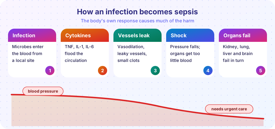
- Recognition. Microbial molecules, such as endotoxin (LPS) of gram-negative bacteria, bind Toll-like receptors on immune cells. Gram-positive bacteria and fungi trigger the same pathways with other molecules. A few toxins act as superantigens (as in toxic shock syndrome) and stimulate huge numbers of T cells at once.
- Cytokine release. Macrophages release TNF, IL-1 and IL-6. These are useful in small amounts and dangerous in large amounts.
- Vessel effects. The vessels dilate (blood pressure falls), capillaries leak (tissue swelling and low blood volume), and the endothelium turns pro-clotting, producing tiny clots in small vessels.
- Shock and organ failure. Tissues receive too little oxygen. The kidney, lung (ARDS), liver, heart and brain fail one after another. Lactate rises from anaerobic metabolism. Septic shock is the situation in which blood pressure cannot be maintained even after fluids and requires drugs to support it.
DIC
In severe sepsis, the clotting system may be activated throughout the body. Clots block small vessels, which damages organs. They use up platelets and clotting factors, leading to bleeding. This is disseminated intravascular coagulation (DIC). A famous example is Waterhouse-Friderichsen syndrome, bleeding into the adrenal glands in meningococcal septicemia, which causes sudden collapse and a skin rash of purple hemorrhages.
Early recognition and urgent treatment improve survival. The key lesson for pathology is that the harm is the consequence of an overwhelming host reaction, not only of the number of microbes.
Common misconceptions
- "A positive TB skin or blood test means active TB." It indicates immune sensitization, which can be latent infection; active disease needs further evidence.
- "Granulomas always mean TB." Many diseases cause them, including sarcoidosis, fungal infection, Crohn disease and foreign material.
- "Sepsis is just a severe infection." It is organ dysfunction from an abnormal response to infection, and can occur even if the original infection looks minor.
- "Antibiotics kill viruses." Antibiotics act on bacteria; viruses need different strategies.
Try it
The bacterial growth plot, the epidemic model and the lookup in the chapter overview are good starting points. This one tracks the TB timeline.
Press Run (or Ctrl + Enter) to see the console output here.
No output.
Check your understanding
Quick check · 5 questions
of 5 answered You scored / 5 · perfect!
-
1. Which cytokine produced by T helper cells activates macrophages in granuloma formation?
Interferon-gamma from Th1 cells activates macrophages and promotes epithelioid and giant cell formation. -
2. The Ghon complex consists of
The Ghon focus plus hilar nodal involvement is the primary TB complex. -
3. Where does secondary (reactivation) TB typically begin?
The apices have high oxygen tension and favor reactivation and cavitation. -
4. Waterhouse-Friderichsen syndrome involves
DIC and sepsis cause adrenal hemorrhage and collapse. -
5. In sepsis, which combination best explains falling blood pressure?
Cytokines dilate vessels and make capillaries leak, lowering effective blood volume.
Summary
- Pus means neutrophils and pyogenic bacteria; lymphocytes and inclusions suggest viruses; granulomas suggest persistent agents such as TB and fungi.
- Tuberculosis causes caseating granulomas; primary disease forms a Ghon complex; reactivation favors the apex and cavities.
- Viruses injure cells directly, through immune attack, or by altering growth.
- Sepsis is organ dysfunction from a dysregulated host response, with cytokines, vasodilation, leakage, DIC and shock.
Next: Stroke and dementia
Track your progress
Create a free account to tick off lessons and pick up where you left off on any device.
of Pathology lessons completed