


















Lesson 14 of 46 · Inflammation and Repair
Healing and Repair: Regeneration, Granulation Tissue and Scars
Learn how tissues heal: regeneration versus scarring, cell types by division capacity, the phases of wound healing, first and second intention, and why healing fails.
Inflammation clears the battlefield; repair rebuilds it. Healing has two possible results. Sometimes the original tissue is restored perfectly, as when the skin's surface renews itself after a graze: this is regeneration. More often, damaged tissue is replaced by a patch of fibrous connective tissue: a scar. A scar holds the tissue together but does not perform the original job. Whether a tissue regenerates or scars depends on the cells that were lost, how big the damage was, and whether the supporting framework survived. This lesson explains the rules and walks through the steps of wound healing.
What you will learn
- Which tissues can regenerate and which can only scar, and why.
- The four overlapping phases of wound healing.
- First and second intention healing.
- The growth factors, cells and factors that control or impair healing.
- Abnormal outcomes: keloids, contracture and fibrosis.
Key terms
Regeneration: replacement of lost cells by the same type, restoring normal structure.
Granulation tissue: soft, pink, highly vascular tissue of new capillaries and fibroblasts that fills a healing wound.
Myofibroblast: a contractile fibroblast that helps close wounds.
Extracellular matrix (ECM): the network of collagen and other proteins that supports cells.
Fibrosis: excessive deposition of collagen and other matrix, a scar-like change in an organ.
Who can regenerate? Labile, stable and permanent cells
Tissues are grouped by how readily their cells divide.
| Group | Behavior | Examples | Result of injury |
|---|---|---|---|
| Labile (continuously dividing) | Constant turnover from stem cells | Surface skin, lining of gut, bone marrow | Usually regenerate completely |
| Stable (quiescent) | Divide only when needed | Liver cells, kidney tubules, endothelium, fibroblasts | Can regenerate if the framework is intact |
| Permanent (non-dividing) | Left the cell cycle for good | Neurons, heart muscle cells; skeletal muscle has limited repair by satellite cells | Replaced by scar (brain: glial scar) |
The liver shows how impressive stable-cell regeneration is. When part of the liver is surgically removed, the remaining cells re-enter the cell cycle and the organ regrows to approximately its original size within weeks to a few months. But regeneration only works if the architecture is preserved. Repeated or severe injury, as with chronic hepatitis or alcohol, leads to scarring and cirrhosis rather than perfect regeneration.
The extracellular matrix is the scaffold. A surviving basement membrane guides regenerating cells into their correct positions. If the scaffold is destroyed, cells regrow in a disordered way, and scar tissue fills the gap. Stem cells also contribute: some tissues contain stem cells (in the base of skin crypts, intestinal crypts and hair follicles) that replace lost cells.
Phases of wound healing
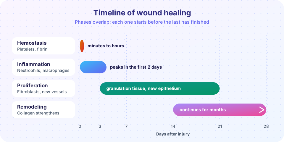
Take a cut skin wound. Four overlapping phases follow, as shown in the timeline above.
- Hemostasis (minutes). Damaged vessels constrict and platelets stick to exposed collagen. A clot of platelets and fibrin plugs the wound and provides a temporary scaffold. Platelets release growth factors such as PDGF and TGF-beta.
- Inflammation (first days). Neutrophils arrive within hours, followed by macrophages over the next two days. They clear bacteria and debris, and the macrophages release growth factors that start the next phase.
- Proliferation (from about day 3 to about week 3). Several things happen in parallel:
- Angiogenesis. New capillaries sprout from existing vessels under the influence of vascular endothelial growth factor (VEGF) and fibroblast growth factor.
- Granulation tissue. The new capillaries and activated fibroblasts form a soft, red, granular-looking tissue (granular because of the many tiny vessels) that fills the defect. It is rich in new vessels and inflammatory cells and poor in collagen at first.
- Re-epithelialization. Cells at the edges of the wound migrate over the granulation tissue and divide until the surface is covered.
- Collagen deposition. Fibroblasts make type III collagen, which is gradually replaced by the stronger type I.
- Wound contraction. Myofibroblasts pull the edges together.
- Remodeling (from about week 2, for months). Granulation tissue matures into a scar. Enzymes called matrix metalloproteinases degrade and rearrange the matrix while fibroblasts lay down more collagen. Collagen is cross-linked, which increases strength. Vessels regress and the scar becomes paler and less cellular.
Strength returns slowly. A skin wound has only about 10 percent of normal strength at the end of the first week. It then increases quickly over the next month or so and approaches, but does not reach, normal: scar tissue usually ends up with about 70 to 80 percent of the strength of unwounded skin.
First and second intention
- Healing by first intention (primary union) applies to a clean wound with edges brought together, such as a surgical incision closed with stitches. The gap is small, little granulation tissue is needed, and the scar is thin.
- Healing by second intention (secondary union) applies to wounds with large tissue loss, such as a deep ulcer or a burn, where the edges cannot be brought together. The defect fills with abundant granulation tissue, contraction is marked, and the scar is larger. Healing takes longer and has more complications.
Inflammation, granulation tissue and scar formation are the same in both. The difference is the amount.
What controls healing
Growth factors and matrix signals coordinate the steps. Epidermal growth factor (EGF) stimulates epithelial cells; VEGF drives new vessels; PDGF and fibroblast growth factor attract and stimulate fibroblasts; TGF-beta is a key promoter of collagen production and scarring. Cell-to-cell contact and ECM signals also tell cells when to stop dividing (contact inhibition).
Why healing fails
- Infection keeps inflammation going and delays repair.
- Poor blood supply, for instance from diabetes or arterial disease, deprives the wound of oxygen and nutrients.
- Nutrition: protein deficiency and lack of vitamin C. Vitamin C is required to hydroxylate proline and lysine in collagen, so deficiency (scurvy) gives weak collagen and poor wound healing. Zinc is also important.
- Glucocorticoids suppress inflammation and collagen production.
- Mechanical factors: movement or tension on the wound, or foreign material in it.
- Diabetes, which impairs circulation, nerves and immune function; diabetic foot ulcers are a classic problem.
Abnormal scarring and fibrosis
- Wound dehiscence (a surgical wound splitting open) and incisional hernia come from weak scar.
- Excess granulation tissue ("proud flesh") protrudes above the wound surface and blocks re-epithelialization.
- Hypertrophic scar and keloid. Both are due to too much collagen. A hypertrophic scar stays within the boundaries of the original wound and often improves; a keloid grows beyond the original wound edges and rarely regresses. Keloids are more common in people with darker skin and tend to run in families.
- Contracture. Excessive contraction, particularly after burns, can deform tissue and limit movement.
- Organ fibrosis. Repeated or persistent injury, with continuing inflammation and TGF-beta signaling, produces scarring inside organs: cirrhosis of the liver, pulmonary fibrosis, kidney scarring, and heart scars after a heart attack. The scar replaces working tissue, reducing function.
Common misconceptions
- "A scar is the same as the original tissue." A scar is made of collagen, has no hair follicles or sweat glands in skin, and lacks the strength and elasticity of the original.
- "Keeping a wound covered and moist delays healing." Moist wound conditions generally help new epithelium migrate. This is a general principle; specific wound care belongs to a clinician.
- "Healing is complete when the skin closes." Remodeling goes on for months or more.
Try it
The first block plots an illustrative curve for wound strength as scar tissue matures. It is an approximate function fitted to the textbook values above, not measured data.
Press Run (or Ctrl + Enter) to see the result here.
Program input · what you'd type while the program runs
Type the answers before you press Run, one per line. Each time the program reads a line (with input()), it gets the next line from this box.
The second block is a small decision helper for first versus second intention healing.
Press Run (or Ctrl + Enter) to see the console output here.
No output.
Check your understanding
Quick check · 6 questions
of 6 answered You scored / 6 · perfect!
-
1. Which cell group can usually regenerate completely after injury, such as the skin's surface?
Labile cells divide continuously from stem cells, so surface epithelia regenerate. -
2. What does granulation tissue consist of?
It is a soft, vascular tissue that fills the wound before collagen matures. -
3. Why does vitamin C deficiency impair healing?
Without vitamin C collagen is weak, as in scurvy. -
4. A scar that grows beyond the boundary of the original wound is called a:
Keloids extend beyond the wound margins; hypertrophic scars stay within them. -
5. Which statement about wound strength is correct?
Strength rises slowly during remodeling but never fully reaches that of unwounded skin. -
6. Which cytokine is a key promoter of collagen production and fibrosis?
TGF-beta drives fibroblasts to make collagen and is central to scarring.
Summary
- Healing is regeneration (restoring the original tissue) or repair (scar).
- Labile and stable cells can regenerate if the scaffold survives; permanent cells are replaced by scar.
- Phases: hemostasis, inflammation, proliferation (angiogenesis, granulation tissue, re-epithelialization, collagen) and remodeling.
- First intention suits clean, approximated wounds; second intention suits wounds with tissue loss.
- Infection, poor perfusion, malnutrition, steroids and diabetes impair healing; excess scarring gives keloids, contractures and organ fibrosis.
Next: Chapter 3 Review
Track your progress
Create a free account to tick off lessons and pick up where you left off on any device.
of Pathology lessons completed