


















Lesson 7 of 46 · Cell Injury and Death
Cellular Adaptations: Hypertrophy, Hyperplasia, Atrophy and Metaplasia
Learn how cells adapt to new demands: hypertrophy, hyperplasia, atrophy and metaplasia, with examples, mechanisms, limits and the link to cancer.
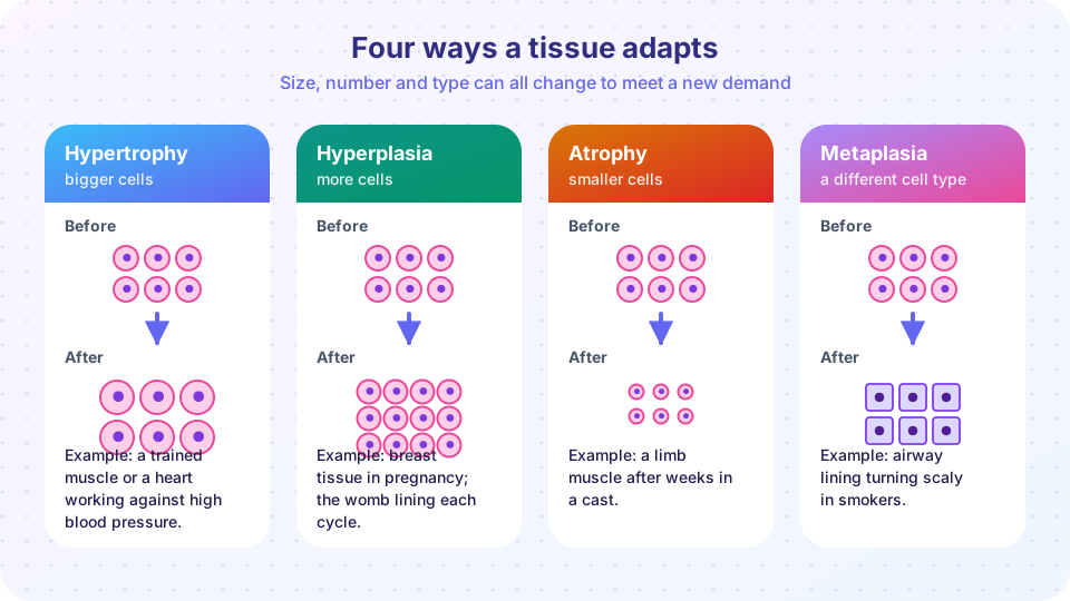
When stress is real but survivable, cells do not simply endure it: they change. They grow, multiply, shrink or switch identity so that the tissue as a whole can cope with its new circumstances. These adaptations are reversible changes in the size, number or type of cells. They are not diseases in themselves, and they can be perfectly normal, as in the muscles of an athlete. But they are also the background to many diseases, from a weakened heart to the earliest steps toward some cancers. Learning to tell them apart on sight is one of the first practical skills in pathology.
What you will learn
- The four adaptive patterns and the one question that separates them.
- Physiologic versus pathologic adaptation, with examples.
- Why heart and skeletal muscle can only enlarge, while liver and skin can multiply.
- The mechanisms behind atrophy and the risks that come with metaplasia.
Key terms
Hypertrophy: increase in the size of cells, giving a bigger organ.
Hyperplasia: increase in the number of cells in an organ or tissue.
Atrophy: decrease in the size of cells (and sometimes the number) after normal growth.
Metaplasia: reversible replacement of one mature cell type by another.
Dysplasia: disordered growth with abnormal-looking cells; not an adaptation but a possible precursor of cancer.
Hypertrophy: bigger cells
In hypertrophy the cells get larger because they make more structural proteins and organelles. The cell number is unchanged. The organ enlarges simply because each cell is bulkier.
Physiologic hypertrophy. Strength training enlarges skeletal muscle fibers. In pregnancy the muscle of the uterus hypertrophies (and also undergoes hyperplasia) under the influence of estrogen.
Pathologic hypertrophy. The classic example is the heart. If blood pressure is chronically high, or a heart valve is narrowed, the left ventricle must push against greater resistance. Its muscle cells respond by adding contractile units, and the wall thickens. This helps at first: a thicker wall can generate more force.
Hypertrophy is the only way to enlarge in cells that cannot divide in adulthood, such as cardiac and skeletal muscle cells. They are called permanent or post-mitotic cells. Hypertrophy has a limit, however. A cell that is too large has blood vessels that cannot keep pace, and the contractile machinery may become inefficient. When the limit is reached, the enlarged heart begins to fail, and some cells die. The adaptation turns into the problem.
A physical reason: wall stress
Why does a thicker wall help? A simplified law of Laplace for a thin-walled sphere says that the stress in the wall is proportional to pressure times radius divided by wall thickness: stress ≈ P × r / (2h). If pressure rises and the wall thickness stays the same, the stress on each muscle fiber rises. Thickening the wall brings the stress back toward normal. The "Try it" below explores this relationship in a simplified form.
Hyperplasia: more cells
Hyperplasia is an increase in cell number. It requires cells that can divide (labile cells like those in skin and gut lining, or stable cells like liver cells that divide when needed). It often occurs together with hypertrophy.
- Hormonal hyperplasia. The female breast grows at puberty and in pregnancy as ducts multiply. The lining of the womb thickens each month under estrogen.
- Compensatory hyperplasia. After a part of the liver is removed, the remaining cells divide and the organ regrows to nearly its original size. The same is true for the remaining kidney after the other is removed, though that is largely hypertrophy.
- Pathologic hyperplasia. It usually results from too much hormonal or growth-factor stimulation. Excess estrogen can cause endometrial hyperplasia, with heavy bleeding. In older men, androgen-driven growth of prostate tissue (benign prostatic hyperplasia) can obstruct the flow of urine.
A key point: hyperplasia is controlled and depends on the stimulus. If the hormone or demand goes away, growth stops and often reverses. This distinguishes it from neoplasia (cancer), where growth continues without the stimulus. Still, an overstimulated, rapidly dividing tissue has more chances to acquire mutations, which is why some pathological hyperplasias, such as endometrial hyperplasia, raise the risk of cancer.
Atrophy: smaller cells
Atrophy is shrinkage of a tissue after it has reached its normal size, because its cells have lost substance. Common causes include:
- decreased workload or disuse (a limb in a cast),
- loss of nerve supply (denervation atrophy),
- diminished blood supply,
- inadequate nutrition,
- loss of hormonal stimulation (the uterus after menopause),
- pressure (a growing tumor can compress and shrink nearby tissue),
- aging.
Atrophic cells are not dead. They have reduced their energy consumption by decreasing protein synthesis and increasing protein breakdown. Degradation happens through two main routes: the ubiquitin-proteasome pathway, where unwanted proteins are tagged and shredded, and autophagy ("self-eating"), where parts of the cell are wrapped in vesicles and digested by lysosomes. Sometimes the cell retains indigestible residue in the form of a brownish pigment called lipofuscin (so-called brown atrophy in the heart and liver of the elderly). If atrophy goes on long enough, cells may eventually die.
Do not confuse atrophy with hypoplasia (an organ that failed to grow fully) or aplasia (an organ that never developed).
Metaplasia: changing identity
In metaplasia one adult cell type is replaced by another adult cell type. It is usually a response to chronic irritation: the new cell type is better able to withstand the stress. Two classic examples:
- Squamous metaplasia in the airways. In long-term smokers, the ciliated, mucus-secreting columnar lining of the bronchi is replaced by flat, tougher squamous cells. The new surface survives smoke better, but the cost is serious: the mucociliary escalator that clears debris is lost.
- Barrett esophagus. Chronic acid reflux changes the normal squamous lining of the lower esophagus into glandular, intestine-like columnar tissue that resists acid better. This is associated with an increased risk of a cancer called adenocarcinoma.
The current understanding is that metaplasia does not occur because a mature cell transforms; rather, local stem cells or progenitors are reprogrammed to differentiate along a new path under altered signals. Because the change is a response to a persistent stimulus, removing the stimulus can sometimes reverse it. If the stimulus persists, the metaplastic epithelium may become dysplastic and, in some cases, cancer may follow. Metaplasia therefore trades function for protection and can be a stage on the path to malignancy.
Common misconceptions
- "Hypertrophy means more cells." No: hypertrophy is larger cells, hyperplasia is more cells. The two often occur together.
- "Metaplasia means the cells are cancerous." Metaplasia is a reversible adaptation. Only some forms are associated with increased cancer risk.
- "Atrophy means the cells are dead." Atrophic cells are alive, smaller and less active.
- "Adaptation is always good." Each adaptation costs something: a thick heart can fail, metaplasia loses function.
Try it
The first block explores the wall stress relationship with made-up relative units. It is a simplified model of a thin-walled ventricle, used only to illustrate why thickening helps.
Press Run (or Ctrl + Enter) to see the result here.
Program input · what you'd type while the program runs
Type the answers before you press Run, one per line. Each time the program reads a line (with input()), it gets the next line from this box.
The second block classifies tissue changes from a change in cell size and cell number. Change the numbers to see the logic.
Press Run (or Ctrl + Enter) to see the console output here.
No output.
Check your understanding
Quick check · 5 questions
of 5 answered You scored / 5 · perfect!
-
1. Which adaptation is the only way for heart muscle cells to enlarge an organ?
Cardiac muscle cells cannot divide in adulthood, so the heart enlarges by growth in cell size. -
2. A smoker's bronchial lining changes from ciliated columnar to squamous cells. This is:
One adult cell type is replaced by another, a reversible response to irritation. -
3. What distinguishes hyperplasia from neoplasia?
Hyperplasia stops or regresses when the stimulus ends; neoplasia is autonomous growth. -
4. Which pathways remove proteins during atrophy?
Atrophic cells increase protein breakdown by tagging proteins for the proteasome and by autophagy in lysosomes. -
5. According to the simplified Laplace relationship, what happens to wall stress if pressure doubles and wall thickness doubles too?
Stress is proportional to pressure divided by thickness; doubling both leaves it unchanged.
Summary
- Adaptations change the size, number or type of cells so a tissue can meet a new demand.
- Hypertrophy is larger cells (limited in muscle that cannot divide); hyperplasia is more cells; atrophy is smaller cells; metaplasia is one cell type replaced by another.
- Hyperplasia is stimulus-dependent and reversible, unlike neoplasia.
- Atrophy reduces energy use through protein breakdown by the proteasome and autophagy.
- Adaptations have costs: heart failure after long hypertrophy, loss of function and cancer risk with some metaplasias.
Track your progress
Create a free account to tick off lessons and pick up where you left off on any device.
of Pathology lessons completed