


















Lesson 27 of 46 · Cardiovascular and Respiratory Pathology
Atherosclerosis and ischemic heart disease
Follow a plaque from fatty streak to rupture, then see how angina and myocardial infarction develop and how heart muscle changes hour by hour after a heart attack.
Atherosclerosis is the most important disease of arteries in the developed world and increasingly everywhere. It is a slow, patchy thickening of the artery wall by fat, cells and scar, and it quietly prepares the ground for the events that people notice: heart attacks, strokes and gangrene of the legs. In the heart it produces ischemic heart disease, a spectrum from brief chest pain to sudden death.
This lesson builds the story in the order it happens in life: first the plaque, then its complications, then the heart's response. Pay attention to why each step follows from the one before it, because that causal chain is what pathologists mean by pathogenesis.
What you will learn
- The layers of a normal artery and how a plaque forms
- The main risk factors and the response-to-injury model
- How plaque rupture causes thrombosis
- The syndromes of ischemic heart disease and their causes
- The time course of tissue change after a myocardial infarction
Key terms
Atherosclerosis: thickening of the arterial intima by a plaque of lipid, inflammatory cells, smooth muscle and connective tissue.
Foam cell: a macrophage swollen with engulfed lipid.
Angina: chest pain caused by reversible myocardial ischemia.
Myocardial infarction (MI): death of heart muscle caused by prolonged ischemia.
Troponin: a heart muscle protein released into blood when myocytes die.
The artery wall and the plaque
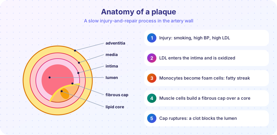
A medium or large artery has three layers. The intima is the innermost, lined by a single sheet of endothelium that keeps blood moving smoothly and releases substances that relax the vessel and prevent clotting. The media is the muscular middle layer, and the adventitia is the outer connective tissue coat. Atherosclerosis happens mainly in the intima.
How a plaque forms
The widely taught response-to-injury hypothesis, developed by Russell Ross, describes a repair process that goes wrong when the injury never stops.
- Endothelial injury or dysfunction. Smoking, high blood pressure, high LDL cholesterol, diabetes and disturbed flow at branch points injure or activate the endothelium, making it more permeable and sticky.
- LDL enters and is modified. Low-density lipoprotein particles slip into the intima, where they are oxidized.
- Inflammation. Monocytes stick to the endothelium, enter the intima, become macrophages and engulf the oxidized LDL. These become foam cells, and a cluster of them is a fatty streak, the earliest visible lesion, which can appear in childhood and is not always destined to progress.
- Smooth muscle migration and scarring. Growth factors released by macrophages, platelets and endothelial cells draw smooth muscle cells from the media into the intima, where they divide and produce collagen. A fibrous cap forms over a growing lipid core that contains dead foam cells, cholesterol crystals and debris.
- Complications. The plaque may calcify, bleed internally, ulcerate or rupture.
A more recent emphasis is on chronic inflammation: plaques are rich in T cells and macrophages, and markers of inflammation predict risk. Lowering LDL cholesterol with statins reduces events, which is why LDL is considered a causal factor, not just a bystander.
Risk factors
Non-modifiable risks include age, male sex (until women's risk catches up after menopause) and family history or genetic predisposition. The most important modifiable risks are hyperlipidemia (especially high LDL and low HDL), hypertension, cigarette smoking and diabetes mellitus. They multiply each other: a person with three risk factors has far more than three times the risk of a person with none. Obesity, physical inactivity and inflammation add to the list.
Where plaques form and what they do
Plaques are most common at the abdominal aorta, the coronary arteries, the popliteal arteries in the legs, the internal carotids and the circle of Willis in the brain. They harm in four ways.
- Stenosis. The plaque narrows the lumen. Because flow depends on radius to the fourth power in a simple tube, a narrowing that is hardly noticeable at rest may fail during exertion.
- Acute plaque change. The cap ruptures, exposing the lipid core and collagen to blood. Platelets stick and the clotting system activates, producing a thrombus that can suddenly block the artery.
- Weakening of the wall. Pressure on the media can destroy it, leading to an aneurysm (a permanent dilation) of the abdominal aorta that can rupture.
- Embolization. Fragments of plaque or clot may break off and block smaller downstream arteries.
Press Run (or Ctrl + Enter) to see the result here.
Program input · what you'd type while the program runs
Type the answers before you press Run, one per line. Each time the program reads a line (with input()), it gets the next line from this box.
Ischemic heart disease
Ischemic heart disease (IHD) is nearly always caused by coronary atherosclerosis. The coronary arteries run on the surface of the heart, and the main ones are the left anterior descending (LAD), the left circumflex and the right coronary artery. The muscle receives most of its blood during diastole, when the heart relaxes, so a fast heart rate shortens the time available for filling the coronary arteries.
The syndromes differ by how complete and how sudden the blockage is.
- Stable angina is chest pressure on exertion that eases with rest. It reflects a fixed, usually large, narrowing: demand exceeds supply, but no cell dies.
- Unstable angina occurs at rest or with a changing pattern. It usually means a partly occluding thrombus on a disrupted plaque. It is dangerous because it may progress to infarction.
- Prinzmetal (variant) angina is caused by coronary spasm.
- Myocardial infarction is necrosis of heart muscle after prolonged ischemia, usually because a plaque ruptured and a thrombus blocked the artery completely.
- Sudden cardiac death is unexpected death from cardiac causes, most often a lethal rhythm disturbance in a person with coronary disease.
- Chronic ischemic heart disease is progressive heart failure from long-standing narrowing and previous infarcts.
Myocardial infarction
The territory depends on the artery. The LAD (the most commonly occluded, in roughly 40 to 50 percent of cases) supplies the front wall and apex of the left ventricle and the front of the septum. The right coronary artery usually supplies the back wall and the back of the septum, and the left circumflex the side wall. A transmural infarction affects the full thickness of the wall and tends to follow a complete blockage; a subendocardial infarction affects the inner layer, which is the most vulnerable because it is farthest from the surface vessels.
Typical presentation includes severe chest pressure, sweating, shortness of breath and nausea, but some patients, especially older adults, women and people with diabetes, have mild or atypical symptoms. Diagnosis rests on symptoms, the electrocardiogram and blood tests for heart muscle proteins, chiefly troponin I or T. Troponin starts to rise within a few hours and stays elevated for about one to two weeks, which makes it sensitive and specific; CK-MB rises and falls faster.
What the muscle looks like over time
Heart muscle dies in a predictable sequence, which pathologists use to date an infarct at autopsy.
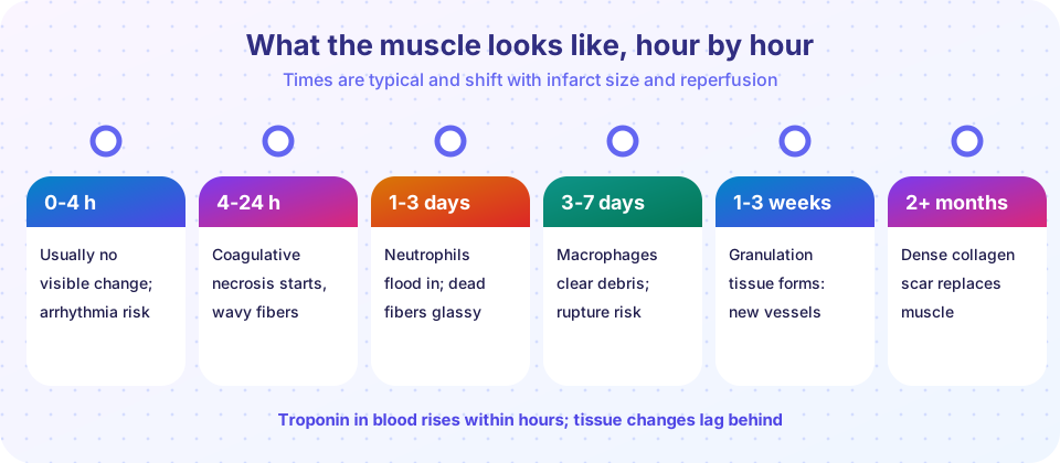
- First 4 hours: little or nothing can be seen under the microscope, even though function is already failing. Electrical instability makes this the period of greatest risk of fatal arrhythmia.
- 4 to 24 hours: coagulative necrosis begins. Cells keep their outline but lose nuclei and look glassy and pink. Contraction bands and waviness of fibers may appear.
- 1 to 3 days: neutrophils invade the dead tissue. Fibers are necrotic and inflamed.
- 3 to 7 days: macrophages remove the debris. The area is soft and yellow, and this is the time when the muscle is weakest, so rupture of the free wall, the septum or a papillary muscle is most likely.
- 1 to 3 weeks: granulation tissue (new capillaries and fibroblasts) replaces the dead tissue.
- After about 2 months: a dense collagen scar remains. Scar does not contract, so a large scar weakens the pump.
Press Run (or Ctrl + Enter) to see the result here.
Program input · what you'd type while the program runs
Type the answers before you press Run, one per line. Each time the program reads a line (with input()), it gets the next line from this box.
Complications
- Arrhythmia is the commonest cause of death in the first hours.
- Pump failure and cardiogenic shock arise when a large part of the muscle is lost.
- Rupture of the free wall (tamponade), septum (shunt) or papillary muscle (acute mitral regurgitation) occurs in the first week.
- Pericarditis produces a rub over the infarct, and a delayed immune form is called Dressler syndrome.
- Mural thrombus forms on dead wall and can embolize, and a weakened scar can bulge as a ventricular aneurysm.
Restoring flow early, by drugs that dissolve the clot or by catheter-based opening of the artery, saves muscle, but it can bring a "reperfusion injury": the returning blood brings oxygen radicals and calcium into stunned cells and can make injury worse for a time.
Common misconceptions
- "A heart attack is a blocked heart." The block is in a coronary artery; the heart muscle downstream dies.
- "Cholesterol builds up like pipe sludge." Plaque is an inflammatory, living lesion in the wall, not just a deposit on it.
- "Only severe narrowing causes heart attacks." Many infarcts begin at plaques that narrowed the lumen only moderately but had thin caps that ruptured.
- "Chest pain is always present." Silent and atypical infarcts are common, especially in diabetes.
Try it
The plots above are teaching models. This small lookup uses the typical time course from the lesson.
Press Run (or Ctrl + Enter) to see the console output here.
No output.
Check your understanding
Quick check · 5 questions
of 5 answered You scored / 5 · perfect!
-
1. What is the first visible lesion of atherosclerosis?
A fatty streak consists of lipid-laden macrophages (foam cells) in the intima. -
2. Which complication is most important in causing acute coronary events?
A ruptured cap exposes the lipid core, platelets and clotting factors form a thrombus, and the artery occludes. -
3. Four to seven days after an infarct, the main risk is related to
At 3 to 7 days macrophages have removed debris and the wall is weakest. -
4. Stable angina usually reflects
Stable angina is reversible ischemia from fixed narrowing. -
5. Which artery is most often involved in myocardial infarction?
The LAD is the most commonly occluded, affecting the front wall, apex and part of the septum.
Summary
- Atherosclerosis is a chronic inflammatory and reparative response in the intima that forms a lipid core under a fibrous cap.
- Risk factors multiply: LDL, hypertension, smoking, diabetes, age and family history.
- Plaque rupture with thrombosis causes most acute events; stable angina reflects fixed narrowing.
- An infarct evolves from coagulative necrosis to neutrophils, macrophages, granulation tissue and scar, with rupture most likely at 3 to 7 days.
- Troponin is the most useful blood marker of heart muscle death.
Track your progress
Create a free account to tick off lessons and pick up where you left off on any device.
of Pathology lessons completed