


















Lesson 17 of 45 · Sensation and Perception
Vision: How the Eye and Brain Create Sight
Learn how the eye focuses light, how rods and cones work, how the brain processes visual information and why trichromatic and opponent-process theories both matter.
Vision is our dominant sense. A large share of the human cortex is devoted to processing what the eyes send, and many everyday phrases ("I see", "it's clear", "in my view") treat sight and understanding as the same thing. Yet the eye is not a camera that delivers a finished picture. It is a living sensor that gathers a patchy, upside-down image, and the brain does astonishing work to turn it into a stable, colorful world.
What you will learn
- What light is and how its physical properties relate to what we see.
- The structure of the eye and how it focuses light.
- How rods and cones differ and why the fovea and blind spot matter.
- How visual information travels and is analyzed in the brain.
- Trichromatic and opponent-process theories of color, and color deficiency.
Key terms
Wavelength: the distance between peaks of a light wave; it corresponds mainly to hue.
Retina: the light-sensitive layer at the back of the eye containing the receptors.
Rods and cones: the two kinds of photoreceptors; rods work in dim light, cones in bright light and give color.
Fovea: a small pit at the center of the retina with the densest cones; it gives the sharpest vision.
Feature detectors: neurons in the visual cortex that respond to particular features such as edges at certain angles.
Light and its properties
Visible light is a narrow band of electromagnetic radiation, with wavelengths from roughly 400 nanometers (violet) to 700 nanometers (red). Three physical properties map onto psychological experience:
| Physical property | What we see |
|---|---|
| Wavelength | Hue (the color name) |
| Amplitude (intensity) | Brightness |
| Purity (mix of wavelengths) | Saturation (vividness) |
Notice that color is not "out there" in the world in the way wavelength is: it is the experience your visual system creates from wavelengths. The same patch of surface can look different in different lighting, and the brain compensates (see perception and illusions).
Structure of the eye
Light passes through these parts in order:
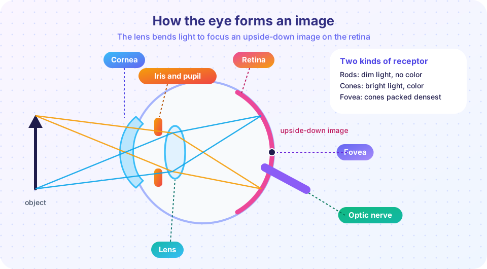
- The cornea, the transparent front window, which does most of the bending of light.
- The pupil, an opening whose size is controlled by the iris (the colored muscle); it widens in dim light and narrows in bright light.
- The lens, which fine-tunes the focus by changing shape (accommodation): thicker for near objects and flatter for far ones.
- The retina, where the image lands, upside down and reversed.
In nearsightedness (myopia), the eyeball is too long or the lens too strong, so distant objects focus in front of the retina and look blurry; in farsightedness, near objects are blurred. Glasses or contact lenses adjust the focus. With age the lens stiffens and near focusing weakens, which is why many people need reading glasses in middle age.
The retina
The retina is a thin layered tissue. Light has to pass through layers of cells to reach the photoreceptors at the back, which absorb it and trigger transduction: a chemical change in light-sensitive pigments that alters the cell's electrical signal. Signals pass to bipolar cells, then to ganglion cells, whose axons bundle together as the optic nerve. Because there are no receptors where the nerve leaves the eye, each eye has a blind spot. You do not notice it because the brain fills in the gap using the surroundings, and the other eye covers it.
Rods and cones
| Rods | Cones | |
|---|---|---|
| Number | About 120 million | About 6 million |
| Best for | Dim light, motion in the periphery | Bright light, color, fine detail |
| Color? | No (shades of gray) | Yes (three types) |
| Location | Mostly outside the fovea | Packed in the fovea |
The numbers are approximate and often quoted in textbooks. In the fovea, cones are densely packed and each connects almost one-to-one with the cells behind it, giving sharp detail. In the periphery many rods share a single path, which makes them sensitive but blurry. At night this causes a curious effect: a faint star is easier to see slightly to the side than directly looked at, because you are using the rods.
When you step from sunlight into a dark theater, you are almost blind for a moment, then gradually see more. This dark adaptation takes about 20 to 30 minutes for rods to reach full sensitivity, because their pigment must regenerate. Red light affects rods less, which is why some night-vision lights are red.
Visual information in the brain
The signals from the optic nerves meet at the optic chiasm, where fibers from the inner half of each retina cross sides. As a result the left visual field of both eyes goes to the right hemisphere and vice versa (the basis of the split-brain demonstration in brain regions). Signals relay in the thalamus and arrive at the primary visual cortex (V1) in the occipital lobe.
In the late 1950s and 1960s David Hubel and Torsten Wiesel recorded from single neurons in the visual cortex of cats and found cells that respond best to a line or edge at a particular angle, a discovery that earned a Nobel Prize in 1981. Such feature detectors are building blocks: later areas combine edges into shapes, faces and scenes.
Beyond V1, processing splits into two main streams. The "what" stream, running toward the temporal lobe, identifies objects and faces. The "where/how" stream, running toward the parietal lobe, handles location and guides movement. Damage can separate them: some patients with injury to the "what" pathway cannot recognize an object but can still reach for it accurately. Others have prosopagnosia (face blindness), where recognizing faces is severely impaired while other objects are recognized normally.
Color vision
Two classic theories seem to compete, but both are right at different stages.
Trichromatic theory (Thomas Young, Hermann von Helmholtz, 1800s). The retina has three kinds of cones, tuned broadly to short ("blue"), medium ("green") and long ("red") wavelengths. All colors come from the pattern of activity across the three. This is the same principle as a screen: red, green and blue lights combine to make other colors.
Opponent-process theory (Ewald Hering, 1800s). Hering noticed that we never see "reddish green" or "yellowish blue" and that after staring at red you see green. He proposed pairs of opposing processes: red against green, blue against yellow, and black against white. Later research found cells in the retina and thalamus that do act as opponents: they are excited by one color and inhibited by its opposite. Staring at a color tires one side of the pair, so when you look away the opposite one dominates, producing an afterimage.
Color deficiency (often called color blindness) usually means a missing or altered cone type. The most common forms affect red-green discrimination, and they are far more common in men, roughly 1 in 12 men of Northern European descent compared with about 1 in 200 women, because the relevant genes lie on the X chromosome. True complete color blindness is rare.
Common misconceptions
"The eye works like a camera." It is a partial analogy at best. The eye has a lens and a light-sensitive surface, but vision depends mostly on the brain's active interpretation.
"We see the image upside down and then the brain flips it." The retinal image is inverted, but the brain does not "flip" it like a photograph; it simply learns to relate retinal positions to the world.
"Color blind people see only black and white." Most have trouble distinguishing particular colors; they still see many colors.
"Carrots give you night vision." Vitamin A is needed for the pigments in the eye, but eating extra carrots will not improve normal night vision. The myth spread from wartime propaganda that concealed the use of radar.
Try it
First, an afterimage demonstration of opponent processing. Press Start, stare at the dot in the center for the full 20 seconds without moving your eyes, then watch the blank panel. Many people see a ghost image in the opposite colors. Experiences vary, and it is not a test of anything.
Second, see additive color mixing, the logic of trichromatic theory. Screens make every color from just red, green and blue lights, as your three cone types respond to different wavelengths.
Third, a schematic of the three cone types across the visible spectrum. The curves are smooth idealized shapes with peaks near commonly cited values, a teaching picture, not measured data.
Press Run (or Ctrl + Enter) to see the result here.
Program input · what you'd type while the program runs
Type the answers before you press Run, one per line. Each time the program reads a line (with input()), it gets the next line from this box.
Check your understanding
Quick check · 6 questions
of 6 answered You scored / 6 · perfect!
-
1. Which physical property of light mostly determines hue?
Wavelength maps to hue; amplitude maps to brightness. -
2. Which receptors work best in dim light but do not provide color?
Rods are very sensitive and mostly outside the fovea; cones need brighter light and give color. -
3. Why does each eye have a blind spot?
That patch has no photoreceptors; the brain fills in the gap. -
4. Afterimages in the opposite color are best explained by
Staring at a color fatigues one half of an opponent pair, so the other half dominates afterwards. -
5. Hubel and Wiesel discovered that cells in the visual cortex
These feature detectors build up shapes and objects from simple features. -
6. Which statement about color deficiency is most accurate?
The common forms involve X-linked genes for the medium and long cones.
Summary
- Light's wavelength, amplitude and purity map to hue, brightness and saturation.
- The cornea and lens focus an inverted image on the retina; the iris controls how much light enters.
- Rods (dim light, no color, periphery) and cones (bright light, color, fovea) transduce light; the optic nerve creates a blind spot.
- Visual cortex neurons detect features; the "what" and "where" streams divide the work.
- Trichromatic theory explains the retina and opponent-process theory explains later stages and afterimages.
Track your progress
Create a free account to tick off lessons and pick up where you left off on any device.
of Introduction to Psychology lessons completed