


















Lesson 13 of 45 · The Brain and Behavior
Brain Regions and Their Jobs
Tour the brain's lobes and deeper structures, meet Phineas Gage and H.M., learn how brain imaging works and why the left-brain right-brain idea is a myth.
If the neuron is the letter, the brain's regions are the words: groups of cells that handle particular kinds of work. Knowing the map helps you make sense of case studies, of what a brain scan can show and of why damage to a small area can have such specific effects. As you read, keep one warning in mind: regions do not work alone. Almost every task, from reading a sentence to riding a bike, uses a network spread across many areas.
What you will learn
- The four lobes of the cerebral cortex and their main functions.
- What the thalamus, hypothalamus, hippocampus, amygdala, basal ganglia, cerebellum and brainstem do.
- What the cases of Phineas Gage and H.M. taught scientists, and how to read them carefully.
- How EEG, CT, MRI, fMRI, PET and TMS work, and what each can and cannot tell you.
- Why "left-brained" and "right-brained" people are a myth.
Key terms
Cerebral cortex: the folded outer layer of the brain, a few millimeters thick, responsible for perception, thinking and voluntary action.
Lateralization: the tendency for some functions to depend more on one hemisphere.
Corpus callosum: a bundle of about 200 million nerve fibers connecting the two hemispheres.
Association areas: regions that combine information from different sources rather than handling one sense or movement.
Plasticity: the brain's ability to change structure and function with experience.
The cerebral cortex and its lobes
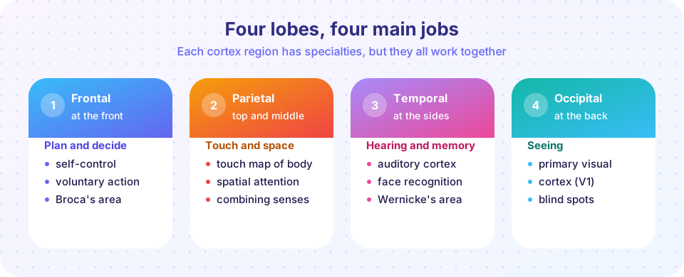
The cortex wraps around the rest of the brain, crumpled into folds that fit a large surface into a small skull. It has two hemispheres, left and right, joined by the corpus callosum. Each hemisphere has four lobes, named for the skull bones above them.
Frontal lobe. At the front. The motor cortex, a strip at its back edge, sends commands to muscles. The prefrontal cortex, in front of that, supports planning, decision making, self-control and working memory. It is also one of the last regions to mature, continuing to develop into the mid-twenties, which is relevant to how adolescents weigh risk and reward (Chapter 8).
Parietal lobe. Behind the central groove. The somatosensory cortex here receives touch, temperature, pressure and pain from the body. The neurosurgeon Wilder Penfield mapped the motor and sensory strips in the 1930s to 1950s by stimulating the exposed brains of patients during surgery; the result is the famous "homunculus", a distorted body map in which sensitive areas such as the lips and fingers take up large areas of cortex.
Occipital lobe. At the back. The primary visual cortex receives signals from the eyes. Damage can cause blind spots even though the eyes are fine.
Temporal lobe. At the sides. It houses the auditory cortex and regions involved in recognizing faces and objects and in memory.
Language areas
In 1861 the French physician Paul Broca examined a patient nicknamed "Tan" who could understand speech but say almost nothing. After his death, Broca found damage in the left frontal lobe, now called Broca's area. In 1874 Carl Wernicke described patients with damage in the left temporal lobe who spoke fluently but meaninglessly and failed to understand speech: Wernicke's area. These discoveries gave early evidence that functions are localized, and modern studies show that language uses a wider network that includes both areas and the fibers linking them.
Beneath the cortex
| Structure | Main roles |
|---|---|
| Thalamus | Relay station: most sensory information passes through it on the way to the cortex |
| Hypothalamus | Hunger, thirst, body temperature, sleep cycles; controls the pituitary gland |
| Hippocampus | Forming new long-term memories; spatial navigation |
| Amygdala | Detecting threat and emotional significance; fear learning |
| Basal ganglia | Starting and smoothing movement; forming habits |
| Cerebellum | Balance, coordination and timing of movement; also involved in some thinking |
| Brainstem (medulla, pons, midbrain) | Breathing, heartbeat, alertness, sleep and waking |
Several of these (hippocampus, amygdala, parts of the hypothalamus and thalamus) are often grouped as the limbic system, which is important for emotion and memory, although scientists dispute exact boundaries. The cerebellum is small in volume but contains most of the brain's neurons.
Two classic cases, read with care
Phineas Gage (1848). A railroad worker in Vermont survived when an iron tamping rod passed through the front of his skull, destroying parts of his left frontal lobe. Early accounts said that his personality changed dramatically. Gage became a symbol of the idea that the frontal lobes shape character. Later historical work shows that many popular details were exaggerated and that Gage's behavior improved over the years, and that he managed to work as a long-distance coach driver in Chile. The lesson is two-sided: the frontal lobes matter for behavior, and a single vivid case is not strong evidence.
H.M. (Henry Molaison). In 1953 surgeons removed parts of his temporal lobes, including most of the hippocampus on both sides, to treat severe epilepsy. Afterwards he could remember his past and talk normally but could not form new lasting memories of events. Yet he could learn new motor skills without remembering the practice sessions. Studied for decades by Brenda Milner and others, H.M. showed that the hippocampus is crucial for forming certain kinds of memory and that memory has more than one system (Chapter 6).
Two hemispheres, one brain
The left hemisphere mostly handles the right side of the body and the right hemisphere the left, because most nerve pathways cross. For most people (nearly all right-handers and a majority of left-handers), language depends mainly on the left hemisphere, while the right plays a larger role in some spatial and attention tasks.
In split-brain research, Roger Sperry and Michael Gazzaniga studied patients whose corpus callosum had been cut to control severe epilepsy. If a word was flashed only to the left visual field (processed by the right hemisphere), the patient could not say what they saw, since the speech-controlling left hemisphere had no access to it, yet the left hand could pick out the matching object. Sperry received a Nobel Prize in 1981.
The myth of "left-brained" and "right-brained" people. Hemispheres specialize in some functions, but healthy brains use both together all the time, through the corpus callosum. A 2013 study that analyzed brain scans of over 1,000 people found no evidence that individuals are stably "left-dominant" or "right-dominant" in their networks. Creativity and logic are not divided between hemispheres.
How we see the living brain
| Method | What it measures | Strengths | Limits |
|---|---|---|---|
| EEG | Electrical activity at the scalp | Millisecond timing, cheap, safe | Poor location of the source |
| CT | X-ray slices of structure | Fast, shows injuries and bleeding | Shows structure, not activity; uses radiation |
| MRI | Detailed structure using magnetic fields | Excellent detail without radiation | Structure, not activity |
| fMRI | Blood-oxygen changes, an indirect sign of activity | Good location (millimeters), safe | Slow (seconds), indirect, expensive |
| PET | Metabolism using a radioactive tracer | Can map chemicals | Uses radiation; slow and costly |
| TMS | Magnetic pulses that briefly disrupt an area | Can test whether an area is necessary | Limited to surface areas; side effects must be screened |
Scans need careful statistics. In a well-known 2009 demonstration, researchers placed a dead salmon in an fMRI scanner, showed it photographs, and, with careless statistics, found apparent "brain activity". The point was not that scans are unreliable, but that analysis must correct for the thousands of comparisons made in each scan.
Common misconceptions
"Each brain area does one thing." Regions have specialties, but most functions rely on networks. Damage to one area often disrupts several abilities.
"fMRI shows thoughts." It shows blood-flow changes, from which researchers infer activity. A bright spot is not a thought and does not prove the area is "the center" of anything.
"Adults cannot change their brains." Learning, practice and recovery all involve plasticity. London taxi drivers who memorized the city's streets showed larger posterior hippocampi on average (Maguire and colleagues, 2000), although such findings do not tell us how much was caused by training.
Try it
First, practice choosing the right brain-imaging method for a question. The answers use the strengths and limits in the table.
Second, a simplified split-brain explainer. Choose where a word is flashed and see what a split-brain patient could say and do. It is a simplified model of the classic findings in typical right-handed patients, for learning only.
Check your understanding
Quick check · 6 questions
of 6 answered You scored / 6 · perfect!
-
1. Which lobe contains the primary visual cortex?
The occipital lobe at the back of the brain processes vision first. -
2. Which structure is crucial for forming new long-term memories?
Cases such as H.M. showed that hippocampal damage prevents new event memories from forming. -
3. Which brain imaging method has the best timing, in milliseconds, but poor location?
EEG records electrical activity at the scalp almost instantly but cannot pinpoint its source. -
4. What is the best summary of the 'left-brain, right-brain' idea?
Large studies do not support stable left-brained or right-brained types. -
5. What does the case of Phineas Gage best illustrate?
Gage's case suggests frontal importance, but later research found popular accounts exaggerated. -
6. The prefrontal cortex is mainly involved in
It supports executive functions and matures late, into the mid-twenties.
Summary
- The cortex has four lobes: frontal (planning, movement), parietal (touch, space), temporal (hearing, memory) and occipital (vision).
- Deeper structures: thalamus (relay), hypothalamus (drives), hippocampus (memory), amygdala (emotion), basal ganglia (movement), cerebellum (coordination) and brainstem (life support).
- Broca and Wernicke linked language to specific areas; Gage and H.M. showed the effects of local damage but need careful reading.
- Imaging methods differ in what they measure and how well; interpretation needs statistics and caution.
- The hemispheres specialize, but people are not "left-brained" or "right-brained".
Track your progress
Create a free account to tick off lessons and pick up where you left off on any device.
of Introduction to Psychology lessons completed