


















Lesson 25 of 45 · Viruses
Viruses: replication cycles
Follow a virus from attachment to release. Compare lytic and lysogenic phage cycles, animal virus entry, one-step growth curves, MOI and plaque assays.
A virus has a simple goal: make more virus. Since it cannot do that alone, replication is a story of hijacking. The virion finds the right cell, gets its genome inside, makes the cell copy that genome and build new coats, assembles the pieces, and sends the new virions out. The steps are the same for nearly all viruses, but the details differ enormously, and those details decide how a virus spreads, which cells it damages and which drugs can stop it.
Bacteriophages are the best way to learn the basics. They were studied first, they grow quickly in the lab, and they gave scientists many of the key ideas of molecular biology. After the phage cycles, you will see how an animal virus differs, and how researchers measure and count viruses even though they are invisible.
What you will learn
- The six general steps of virus replication
- How the lytic and lysogenic cycles differ and what triggers induction
- How animal viruses enter cells and acquire envelopes
- How to read a one-step growth curve and what burst size means
- How to calculate multiplicity of infection and plaque assay titers
Key terms
Lytic cycle: replication that ends with the host cell bursting and releasing new virions.
Lysogeny: a state in which phage DNA is integrated into the host chromosome and copied with it.
Prophage: the integrated phage genome.
Burst size: the average number of new virions released from one infected cell.
MOI: multiplicity of infection, the ratio of virus particles to host cells.
Six steps that most viruses share
- Attachment. A virion binds a specific receptor on the host cell surface. This is why viruses have a narrow host range. Influenza's hemagglutinin binds sialic acid on airway cells, SARS-CoV-2's spike binds the protein ACE2, and HIV binds CD4 and a co-receptor on certain immune cells.
- Entry. Phages inject their genome through the cell wall and leave the empty capsid outside. Enveloped animal viruses either fuse with the cell membrane or are taken in by endocytosis, then fuse from within the vesicle. Non-enveloped animal viruses are usually taken in by endocytosis and escape from the vesicle.
- Uncoating. The capsid is opened or removed to release the genome so the cell can read it.
- Synthesis. The genome is copied and its genes are expressed. In the Baltimore scheme the exact path to mRNA depends on the genome type. Early genes usually make enzymes for copying, late genes make the structural proteins.
- Assembly. Coat proteins and genomes come together into new virions, sometimes with help from host chaperones.
- Release. New virions leave by lysis, which kills the cell, or by budding through the membrane, which can let the cell stay alive for a while and acquires the envelope on the way out.
The lytic cycle of a phage
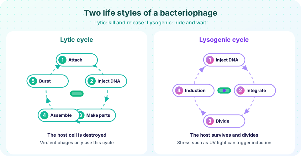
Consider T4 phage infecting E. coli at 37 °C. The tail fibers attach to the outer membrane, the sheath contracts and the DNA is injected within a minute or two. Phage enzymes quickly take over: they degrade the host chromosome into nucleotides that are re-used for phage DNA, and they switch the host's RNA polymerase to read phage genes. For about the first ten to fifteen minutes you cannot find any complete virions inside the cell, because the phage has been taken apart into its genome and parts. This quiet interval is the eclipse period. Then new heads and tails assemble, the DNA is packed into heads, and tails are attached. A phage enzyme, lysozyme, finally digests the cell wall, the cell bursts and releases typically a hundred or more phages. A complete cycle takes roughly 25 to 30 minutes under good conditions. The time from adsorption to the first release is the latent period. Phages that use only this cycle are called virulent.
The lysogenic cycle
Some phages, called temperate, have a second option. After injection, the phage DNA may circularize and integrate into the host chromosome using an enzyme called integrase. The integrated genome is a prophage. Phage lambda is the classic example. The host, now a lysogen, grows and divides normally, and each daughter inherits a copy of the prophage. A repressor protein made from the prophage switches off the lytic genes and also makes the cell immune to a second infection by the same phage.
The decision between the lytic and lysogenic paths depends on conditions in the host: in a starving or crowded cell the lysogenic path is more likely. The prophage can be induced later. Damage to the host's DNA, for example from ultraviolet light, triggers the cell's SOS response, in which a protein called RecA cuts the repressor. Then the lytic genes switch on, the prophage leaves the chromosome and the cell makes and releases phages. Induction is, from the phage's point of view, an escape from a sinking ship.
Lysogeny matters for human disease. Several bacterial toxins are encoded by prophages: the diphtheria toxin, the cholera toxin and the Shiga toxin of E. coli O157:H7 are examples. A harmless bacterium can turn dangerous when it becomes infected by the right phage. This is lysogenic conversion.
How animal viruses differ
Most animal viruses do not inject their genome. They are taken up whole, and the cell's compartments are used for uncoating. DNA viruses, such as adenoviruses, usually replicate in the nucleus, where the host DNA machinery is, while most RNA viruses replicate in the cytoplasm. Poxviruses are a DNA exception: they carry their own enzymes and replicate in the cytoplasm. Release by budding gives the envelope. Some animal viruses can establish latency, resembling lysogeny: herpes simplex virus hides in sensory neurons for life and may reactivate to cause cold sores. Retroviruses such as HIV integrate a DNA copy of their genome into a host chromosome as a provirus, where it is invisible to the immune system until it is expressed.
Because every virus relies on the host, antiviral drugs are hard to design, but they exist. Acyclovir blocks herpes virus DNA synthesis and is activated only in infected cells. Oseltamivir inhibits influenza neuraminidase and so blocks the release of virions. Antiretroviral drugs block HIV reverse transcriptase, integrase or protease. Our body also has antiviral defenses, such as interferons, signal proteins released by infected cells that warn neighbors to prepare.
Measuring phages
Scientists count viruses by what they do. In a one-step growth experiment, a culture is infected with a high MOI, then diluted so that new infections are rare. Samples are taken at intervals and the number of infectious phages is counted. The curve has a flat early part (during the eclipse and latent period the count of free phage stays low), a sharp rise when the cells burst and a plateau. The burst size is the plateau titer minus the starting titer, divided by the number of infected cells.
Phage particles can also be counted on a plaque assay. A sample is diluted in steps of ten, mixed with bacteria and spread in soft agar on a plate. Each infectious phage infects a bacterium, multiplies, spreads to neighbors and leaves a clear circle, a plaque, in the bacterial lawn. One plaque represents one plaque-forming unit (PFU). The concentration is the number of plaques divided by the dilution and the volume plated. Plates with 30 to 300 plaques are the most reliable to count.
The multiplicity of infection (MOI) is the ratio of virions to cells. It follows a Poisson distribution: with an average of m virions per cell, the fraction of cells receiving exactly k virions is e^(−m) x m^k / k!. At an MOI of 1, about 37% of the cells (the e^(−1) fraction) get no virion at all, which surprises many people. To infect nearly every cell you need an MOI of 5 or more.
Common misconceptions
"A virus eats the cell." Viruses do not digest anything. They redirect the cell's own machinery. A cell may die in the end, but only after it has been busy building virions.
"Integrated means harmless." A prophage can carry genes that make the host more dangerous, and latent viruses can reactivate.
"An MOI of 1 infects every cell." It does not. By chance, about 37% of cells receive no virus at all and about 26% receive two or more.
Try it
The first demonstration models a one-step growth curve. Change the latent period or burst size and watch how the curve moves. The model is a simple illustration: cells burst at times that follow a normal distribution.
Press Run (or Ctrl + Enter) to see the result here.
Program input · what you'd type while the program runs
Type the answers before you press Run, one per line. Each time the program reads a line (with input()), it gets the next line from this box.
The second demonstration shows how the Poisson formula divides cells at different MOI values.
The third demonstration turns plaque counts into a titer. Try a different count or dilution.
Press Run (or Ctrl + Enter) to see the result here.
Program input · what you'd type while the program runs
Type the answers before you press Run, one per line. Each time the program reads a line (with input()), it gets the next line from this box.
Check your understanding
Quick check · 6 questions
of 6 answered You scored / 6 · perfect!
-
1. What is the eclipse period of a lytic phage infection?
After injection the phage genome is taking over the cell and making parts, so no assembled infectious phages exist yet. -
2. What is a prophage?
A prophage is the integrated form of a temperate phage and is copied along with the host DNA. -
3. Which event commonly triggers induction of a prophage?
DNA damage triggers the SOS response, RecA cleaves the repressor and the lytic genes switch on. -
4. On a plaque assay plate, what does a single plaque represent?
Each plaque grows from a single infectious particle that infected a cell and spread to its neighbors. -
5. At an MOI of 1, roughly what fraction of cells receives no virion at all?
The Poisson probability of zero is e to the power of minus 1, about 0.37. -
6. How can a harmless bacterium become a toxin producer through a phage?
Diphtheria, cholera and Shiga toxin genes are carried by prophages.
Summary
- Replication follows six steps: attachment, entry, uncoating, synthesis, assembly and release.
- Receptor binding explains a virus's narrow host and tissue range.
- Virulent phages use the lytic cycle; temperate phages can instead integrate as a prophage and wait for induction.
- Prophages can add toxin genes to the host (lysogenic conversion).
- Animal viruses are taken in whole, can bud with an envelope, and may establish latency or a provirus.
- Phage titers are counted by plaque assays; MOI follows a Poisson distribution.
Next: Chapter 5 review
Track your progress
Create a free account to tick off lessons and pick up where you left off on any device.
of Microbiology lessons completed