


















Lesson 5 of 45 · Getting Started
Chapter 1 Review: Getting Started
Review the history of microbiology, microscopy, stains and culture methods from Chapter 1 with a recap table, flashcards and a ten-question quiz.
You now have the toolkit that every later chapter relies on. You know how microbes were discovered and how experiments tied them to disease, you can judge what a microscope can and cannot show, you understand the logic of the Gram stain, and you can turn a murky culture into a number of colony-forming units. This review pulls the chapter into one page, gives you a way to practice recall, and ends with a longer quiz.
What you will learn
- Which ideas from Chapter 1 you have mastered and which need another look.
- How the four lessons connect to each other.
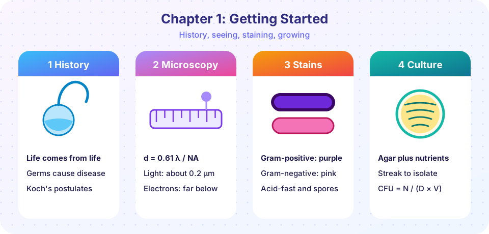
The chapter at a glance
| Lesson | Big idea | Formulas and facts to remember |
|---|---|---|
| History | Experiments, not opinions, built microbiology | Leeuwenhoek 1670s; Pasteur's swan-neck flasks 1861; Koch's four postulates; Jenner 1796; Fleming 1928; Woese 1977 |
| Microscopy | Resolution limits what you can see | total magnification = objective × eyepiece; d = 0.61 λ / NA; light about 0.2 µm; 1 mm = 1,000 µm = 1,000,000 nm |
| Stains and media | Stains add contrast and sort cells; agar gives pure cultures | Gram: crystal violet, iodine, alcohol, safranin; CFU/mL = colonies / (dilution × volume); count 30 to 300 |
How the lessons connect
The history lesson explains why we use pure cultures: Koch needed one kind of bacterium at a time to prove it caused a disease. The microscopy lesson explains why stains are needed: living cells give little contrast, and they are near the limit of resolution anyway. The staining lesson explains why we plate and count: once a colony is isolated and Gram-stained, it can be identified, grown and studied. Each tool covers the weakness of the one before it.
Common mistakes to avoid
Students who lose marks on this chapter usually do so in one of four places.
- Mixing up units. A micrometer is 1,000 times larger than a nanometer. Write the unit in every answer and convert before you compare sizes. A quick check: a bacterium (about 2 µm) should always come out bigger than a virus (about 0.1 µm).
- Confusing magnification with resolution. If a question asks what limits the detail you can see, the answer is resolution (wavelength and numerical aperture), not the power of the lenses.
- Forgetting the order of the Gram stain. The sequence is crystal violet, iodine, alcohol, safranin. Remember that the alcohol step is the one that makes the difference between the two groups.
- Counting the wrong plate. Only plates with 30 to 300 colonies are used, and the answer must be divided by both the dilution and the volume plated.
Study checklist
Before moving on, make sure you can do each of these without looking at the lessons: name two experiments that refuted spontaneous generation or supported the germ theory; calculate total magnification and field of view; use d = 0.61 λ / NA for one wavelength and one numerical aperture; describe what each Gram stain reagent does; and turn a plate count into CFU per milliliter. If one of these feels shaky, reread the matching lesson from the table above and then try the flashcards again.
Practice with flashcards
Recall is the most effective way to study. Flip each card, say the answer aloud first, then check.
Review quiz
Quick check · 10 questions
of 10 answered You scored / 10 · perfect!
-
1. Which discovery is credited to Antonie van Leeuwenhoek?
Leeuwenhoek described 'animalcules' and bacteria in the 1670s and 1680s. Koch proved germ theory, Petri designed the dish and Fleming noticed penicillin. -
2. What was the key variable Pasteur changed in his flask experiment?
Only the neck shape differed, so contamination could be traced to airborne particles being trapped in the bend. -
3. A virus of 80 nm is how many micrometers across?
Divide nanometers by 1,000: 80 / 1,000 = 0.08 µm. -
4. Total magnification with a 100× objective and 10× eyepiece is...
Multiply objective and eyepiece: 100 × 10 = 1,000×. -
5. Using the formula d = 0.61 λ / NA with λ = 500 nm and NA = 1.0, what is d?
0.61 × 500 / 1.0 = 305 nm. -
6. After the alcohol step of the Gram stain, which cells are colorless?
Gram-positive cells keep the dye in their thick wall; Gram-negative cells lose it and then take up safranin. -
7. Why might a scientist choose phase-contrast microscopy?
Phase-contrast converts small differences in light speed through the cell into brightness differences. -
8. A 0.1 mL sample of the 10⁻⁶ dilution gives 45 colonies. How many CFU/mL were in the original?
45 / (10⁻⁶ × 0.1) = 45 / 10⁻⁷ = 4.5 × 10⁸ CFU/mL. -
9. Which of Koch's postulates is hardest to meet for a virus?
Viruses need living host cells, so a pure culture on lifeless medium is impossible. -
10. Which sterilization method kills bacterial endospores in a laboratory?
Steam under pressure at 121 °C for 15 minutes or longer destroys spores; alcohol and cold do not.
Summary
- Chapter 1 gave you the history, the optics, the stains and the culture methods that microbiology rests on.
- The tools support each other: pure cultures need media, identification needs stains, and stains need good microscopy.
- You can now calculate magnification, resolution limits and CFU counts.
Track your progress
Create a free account to tick off lessons and pick up where you left off on any device.
of Microbiology lessons completed