


















Lesson 3 of 45 · Getting Started
Microscopy and the Scale of Life
Learn how light and electron microscopes work, why resolution matters more than magnification, and how to calculate sizes from micrometers to nanometers.
A microscope does two jobs, and beginners usually remember only one. The first is to magnify: to make the image bigger. The second, which matters far more, is to resolve: to show two nearby points as two points instead of one blur. You can enlarge a blurry photograph as much as you like and it will never show new detail. The same is true of a microscope, and that is why microbiologists talk about resolution before they talk about power.
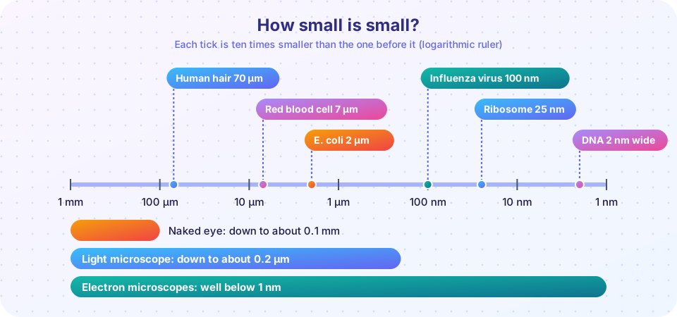
This lesson gives you a working feel for size. You will convert between millimeters, micrometers and nanometers, calculate total magnification and field of view, use the formula that sets the limit of light microscopy, and compare the main kinds of microscope that a microbiologist might choose.
What you will learn
- How to convert between mm, µm and nm without mistakes.
- The difference between magnification, resolution and contrast.
- How to use
d = 0.61 × λ / NAto find the smallest detail a lens can resolve. - When to choose bright-field, phase-contrast, dark-field, fluorescence, confocal, TEM or SEM.
Key terms
Magnification: how many times larger the image looks than the object.
Resolution (resolving power): the smallest distance at which two points are still seen as separate.
Numerical aperture (NA): a number describing how much light a lens collects; larger NA means finer resolution.
Contrast: the difference in brightness or color between an object and its surroundings.
Wavelength (λ): the distance between wave crests; light has a wavelength of roughly 400 to 700 nm.
Units you must know
Microbiology uses three length units constantly.
| Unit | Symbol | In meters | Relationship |
|---|---|---|---|
| millimeter | mm | 0.001 m | 1 mm = 1,000 µm |
| micrometer | µm | 0.000001 m | 1 µm = 1,000 nm |
| nanometer | nm | 0.000000001 m | 1 nm = 0.001 µm |
Convert down by multiplying by 1,000 for each step (mm to µm to nm), and up by dividing. A virus of 120 nm is 0.12 µm. A bacterium of 3 µm is 3,000 nm. Always write the unit; a bare "3" is meaningless in this subject.
Magnification: easy to calculate
A compound light microscope has two sets of lenses. The objective near the specimen (usually 4×, 10×, 40× or 100×) forms an enlarged image, and the eyepiece (ocular, normally 10×) magnifies it again. Total magnification is the product:
total magnification = objective × eyepiece
So a 40× objective with a 10× eyepiece gives 400×, and the 100× objective gives 1,000×. The field of view (how much of the slide you see) shrinks as magnification rises. A standard eyepiece has a field number of about 18 mm, so the diameter of the area you see is roughly field number ÷ objective magnification. At 100× that is 18 ÷ 100 = 0.18 mm, or 180 µm. About ninety E. coli cells (2 µm each) placed end to end would cross it.
Resolution: the real limit
Light behaves as a wave. When it passes through a tiny object it spreads out (diffracts), so a point of light becomes a small disc. Two points closer than about the width of that disc merge. Ernst Abbe worked this out in the 1870s, and the version used in textbooks is:
d = 0.61 × λ / NA
where d is the smallest resolvable distance, λ is the wavelength of light, and NA is the numerical aperture of the objective. NA is written on the lens barrel. For a dry lens it cannot exceed 1, because it equals n × sin θ and air has a refractive index n of 1. An oil-immersion objective fills the gap between slide and lens with oil of refractive index about 1.5, matching glass, so more light is captured and NA can reach about 1.4.
Worked example: green light (λ = 550 nm) with an oil objective of NA 1.4 gives d = 0.61 × 550 / 1.4, about 240 nm, or 0.24 µm. This is why the light microscope tops out at around 0.2 µm. Shorter (blue) wavelengths help a little, but not enough to see a 100 nm virus particle as anything but a faint dot.
Magnification beyond what resolution supports is called empty magnification: the picture gets bigger but no clearer.
Press Run (or Ctrl + Enter) to see the result here.
Program input · what you'd type while the program runs
Type the answers before you press Run, one per line. Each time the program reads a line (with input()), it gets the next line from this box.
Contrast: seeing what is nearly transparent
Most live microbes are almost colorless and watery, so they hardly change the light and are hard to see. Microscopists solve this by adding contrast:
- Bright-field microscopy lights the sample from below and sees dark or stained objects on a bright background. It is the standard method, normally with stains (next lesson).
- Dark-field microscopy blocks the direct light so only light scattered by the sample reaches the lens. Live cells glow on a black background, useful for thin spirochetes.
- Phase-contrast microscopy, invented by Frits Zernike (Nobel Prize, 1953), turns tiny differences in how much a structure slows light into differences in brightness. It shows live, unstained cells and their internal structures.
- Fluorescence microscopy excites dyes or proteins (such as green fluorescent protein) with short-wavelength light and collects the longer-wavelength glow. Labeled antibodies can light up one species or one protein.
- Confocal microscopy adds a pinhole that blocks out-of-focus light, building sharp optical slices that can be stacked into a 3D image, for example of a biofilm.
Electron microscopes
Electrons accelerated through 100 kV have a wavelength of only about 4 picometers, more than a hundred thousand times shorter than visible light, so in principle they resolve atoms; in practice lens errors limit biological images to roughly nanometers and below. The specimen sits in a vacuum, so everything seen is dead and prepared.
- Transmission electron microscopy (TEM) sends electrons through an extremely thin sample and shows internal structure: ribosomes, the layers of a cell envelope, the inside of a virus particle. Cryo-electron microscopy, which freezes samples so rapidly that the water does not form crystals, earned the 2017 Nobel Prize in Chemistry.
- Scanning electron microscopy (SEM) sweeps a beam over a coated surface and builds a 3D-looking picture of its shape, such as the surface of a bacterial colony or a pollen grain.
Common misconceptions
"Higher magnification always means a better look." Without more resolution you only enlarge the blur. Always check NA and wavelength first.
"You can see viruses with a school microscope." Most viruses are smaller than the 0.2 µm limit, so you can see them only with an electron microscope (or detect their effects, such as plaques in a bacterial lawn).
"Electron micrographs show true color." They are monochrome images; any color is added later by software to help the reader.
Check your understanding
Quick check · 5 questions
of 5 answered You scored / 5 · perfect!
-
1. A student uses a 10× eyepiece and a 40× objective. What is the total magnification?
Total magnification is the product of the lenses: 10 × 40 = 400×. -
2. Convert 450 nm to micrometers.
Divide by 1,000: 450 nm = 0.45 µm. -
3. Which change makes a light microscope resolve smaller details?
In d = 0.61 λ / NA, resolution improves when NA rises or λ falls. Red light has the longer wavelength, so it is worse than blue. -
4. Which technique would you pick to see the 3D surface shape of a bacterial colony at high magnification?
SEM scans a beam over the surface and gives a three-dimensional-looking image. TEM shows internal slices. -
5. Why is a drop of oil placed between the slide and the 100× objective?
Oil with a refractive index close to glass stops light bending away from the lens, so NA (and therefore resolution) increases. The field of view actually shrinks at 100×.
Summary
- Resolution, not magnification, limits what you can see. The light microscope resolves down to about 0.2 µm.
- Total magnification = objective × eyepiece; field of view = field number ÷ objective power.
d = 0.61 × λ / NA: shorter wavelengths and larger NA give finer resolution. Oil immersion raises NA.- Contrast methods (dark-field, phase-contrast, fluorescence, confocal) let you see live, unstained cells.
- Electron microscopes (TEM for interiors, SEM for surfaces) reach the scale of viruses and molecules but only for dead, prepared samples.
Track your progress
Create a free account to tick off lessons and pick up where you left off on any device.
of Microbiology lessons completed