


















Lesson 12 of 45 · Bacterial Structure and Growth
The Bacterial Cell Envelope
Understand the plasma membrane, peptidoglycan wall, Gram-positive and Gram-negative envelopes, LPS endotoxin, and why penicillin and lysozyme kill bacteria.
Imagine a balloon filled with a thick sugar solution and dropped into fresh water. Water rushes in, the balloon swells and bursts. That is the permanent danger every bacterial cell faces, because the cytoplasm is a far more concentrated solution than most places bacteria live. The cell envelope is the engineering answer: a membrane that controls what enters, and a wall that is strong enough to hold the pressure.
The envelope is also the single most important target in medicine. Penicillin, the first widely used antibiotic, attacks it. Lysozyme in your tears and saliva attacks it. The Gram stain you met in Chapter 1 is really a test of how the envelope is built. This lesson walks through the layers from the inside out and shows why the two main designs behave so differently.
What you will learn
- How the plasma membrane works and what it does in bacteria.
- How peptidoglycan is built and why cross-links matter.
- The structural differences between Gram-positive and Gram-negative envelopes.
- Why lipopolysaccharide (endotoxin) matters in disease.
- How penicillin, lysozyme and osmosis relate to the wall.
Key terms
Peptidoglycan (murein): a net of long sugar chains joined by short peptide bridges; unique to bacteria.
Teichoic acids: polymers in the Gram-positive wall that help hold it together and bind ions.
Lipopolysaccharide (LPS): a large molecule in the outer leaflet of the Gram-negative outer membrane; the lipid part (lipid A) is an endotoxin.
Periplasm: the gel-like space between the inner and outer membranes of Gram-negative bacteria.
Osmosis: movement of water across a membrane toward the side with more dissolved solute.
The plasma membrane
Under the wall sits the plasma membrane, a double layer of phospholipids with embedded proteins. The fatty tails point inward and the charged heads face the water on both sides, so the membrane is a barrier to most dissolved substances. Proteins in it do three big jobs: they move nutrients and ions in and out (by diffusion, carrier proteins, or pumps that spend energy), they run the electron transport chain, and they hold ATP synthase.
That second job is a notable difference from eukaryotes. Your cells carry out respiration in mitochondria. A bacterium has no mitochondria, so it uses its own plasma membrane: electron carriers pump protons out, a gradient forms, and protons flow back through ATP synthase to make ATP. The membrane is the cell's power station as well as its border, and Chapter 4 returns to this.
Bacterial membranes mostly lack cholesterol and other sterols (some use related molecules called hopanoids to keep the membrane stable).
Peptidoglycan: a net around the cell
Outside the membrane lies the cell wall, made of peptidoglycan. Its building blocks are two sugars, N-acetylglucosamine (NAG) and N-acetylmuramic acid (NAM), which alternate to form long chains. Each NAM carries a short chain of amino acids (a peptide), including unusual D-forms of amino acids that most other organisms do not use. Neighboring chains are connected by cross-links between these peptides, like the rungs of a ladder or the weave of a fishing net. The result is one giant molecule wrapped around the whole cell, which is why it is also called the sacculus.
The cross-links are made by enzymes called transpeptidases, also known as penicillin-binding proteins. Cells that grow must keep making new peptidoglycan. Special enzymes cut small gaps in the old net, new strands are inserted and cross-linked, and the cell lengthens.
Gram-positive and Gram-negative designs
| Feature | Gram-positive | Gram-negative |
|---|---|---|
| Peptidoglycan | thick (roughly 20 to 80 nm), many layers | thin (a few nm), one or few layers |
| Outer membrane | absent | present, with LPS |
| Teichoic acids | present | absent |
| Periplasm | narrow | wide, with enzymes |
| Susceptibility to lysozyme | high | lower (outer membrane protects) |
| Typical examples | Staphylococcus, Bacillus, Streptococcus | E. coli, Salmonella, Pseudomonas |
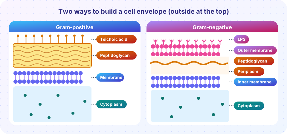
In the Gram-positive envelope, the plasma membrane is covered by a thick peptidoglycan wall threaded with teichoic acids, polymers that help to hold the wall together and move ions. This thick layer is what traps the crystal violet-iodine complex in the Gram stain.
In the Gram-negative envelope, the peptidoglycan is thin and sits in the periplasm, between the inner (plasma) membrane and an outer membrane. The outer membrane's inner leaflet is made of ordinary phospholipids, but its outer leaflet is made of LPS. Channel proteins called porins let small molecules through. This membrane is a strong barrier: it keeps out many antibiotics, bile salts and detergents, which explains why Gram-negative infections are often harder to treat.
LPS has three parts: a lipid anchor (lipid A), a core sugar chain and a long variable sugar chain (the O antigen) that points outward. Lipid A is an endotoxin. When Gram-negative bacteria die and break up, lipid A is released and the immune system reacts strongly, causing fever, a drop in blood pressure and, in severe infections, septic shock. That is why killing the bacteria with an antibiotic can occasionally make a patient feel worse before better.
Other envelopes
- Acid-fast bacteria (Mycobacterium) have a Gram-positive-like wall coated with waxy mycolic acids, which make them tough, slow-growing and resistant to many drugs and stains.
- Mycoplasma has no cell wall at all, so penicillin has no effect on it. Its membrane is strengthened with sterols taken from the host.
- Archaea have no peptidoglycan. Many have a surface protein layer (an S-layer), and some methanogens have a wall of a related polymer called pseudomurein.
Osmosis and the wall
The cytoplasm of a bacterium holds a high concentration of dissolved solutes. In dilute surroundings water enters and creates turgor pressure on the wall, on the order of a few atmospheres for E. coli and higher for some Gram-positives. The wall prevents bursting. In a concentrated solution (salty or sugary), the opposite happens: water leaves and the membrane shrinks away from the wall (plasmolysis). This is why salting and sugaring preserve food (see Growth Conditions and Control).
The van 't Hoff relation gives the osmotic pressure of a solution: Π = C × R × T, where C is the total concentration of dissolved particles in mol/L, R = 0.08206 L·atm/(mol·K) and T is the temperature in kelvin. The calculator below uses an assumed internal concentration; real values vary by species and conditions.
Press Run (or Ctrl + Enter) to see the result here.
Program input · what you'd type while the program runs
Type the answers before you press Run, one per line. Each time the program reads a line (with input()), it gets the next line from this box.
Penicillin and lysozyme
Lysozyme is an enzyme found in tears, saliva and egg white. Alexander Fleming noticed it in 1921, before penicillin. It cuts the bond between NAM and NAG in the peptidoglycan chain, so the sugar backbone falls apart. It works well on Gram-positive bacteria, whose wall is exposed, and less well on Gram-negative cells whose wall is protected by the outer membrane.
Penicillin and its relatives (the beta-lactam antibiotics) block the transpeptidases that build the cross-links. Growing cells keep cutting the old wall and inserting new strands, but the new strands are not cross-linked, so the wall becomes weak at exactly the places where it grows. Water pressure then bursts the cell. Because human cells have no peptidoglycan, penicillin is largely harmless to us. It only kills growing cells; resting cells survive.
Common misconceptions
"Gram-negative bacteria have no cell wall." They do; it is just thin and sits between two membranes.
"Penicillin works on all bacteria." It does not work on Mycoplasma (no wall), and many Gram-negative bacteria are protected by the outer membrane or destroy the drug with enzymes (beta-lactamases).
"Gram-negative bacteria are more dangerous." Danger depends on the species; but LPS endotoxin and outer-membrane barriers make Gram-negative infections especially difficult.
Check your understanding
Quick check · 5 questions
of 5 answered You scored / 5 · perfect!
-
1. Which polymer forms the bacterial cell wall?
Peptidoglycan, a net of sugar chains joined by peptide cross-links, is unique to bacteria. -
2. What does penicillin block?
Penicillin inhibits the transpeptidases that cross-link the wall, so growing cells burst. -
3. Which part of Gram-negative bacteria contains lipid A (endotoxin)?
Lipid A anchors LPS in the outer membrane; when released it triggers fever and shock. -
4. A cell with an internal solute concentration of 0.40 mol/L sits in pure water at 37 °C. The net outward pressure is approximately...
Π = 0.40 × 0.08206 × 310 ≈ 10.2 atm. The wall must withstand that pressure. -
5. Why is Mycoplasma unaffected by penicillin?
Penicillin targets wall synthesis; Mycoplasma has no wall to target.
Summary
- The plasma membrane controls transport and, in bacteria, also hosts the respiratory chain and ATP synthase.
- Peptidoglycan is a cross-linked sugar-peptide net that holds back turgor pressure.
- Gram-positive: thick wall plus teichoic acids. Gram-negative: thin wall between an inner and an outer membrane that carries LPS.
- Lipid A of LPS is an endotoxin; the outer membrane makes Gram-negative bacteria harder to treat.
- Penicillin blocks cross-linking; lysozyme cuts the sugar chains.
Track your progress
Create a free account to tick off lessons and pick up where you left off on any device.
of Microbiology lessons completed